Xolography is a novel light printing technique that has been explored for dental products and in-space manufacturing. At TU/e, this technique has been adapted to 3D print living cells. This research can pave the way for 3D printed kidneys and muscle tissue. The team pioneered the Xolography-based method to produce tiny structures with features as small as 20 µm-approximately the size of a human cell. These results were published in Advanced Materials.
Is Xolography the technique that will enable a future of 3D-printed hearts and kidneys? "Unfortunately, this is still entirely speculative for now, I'm afraid," Miguel Dias Castilho tempers expectations.
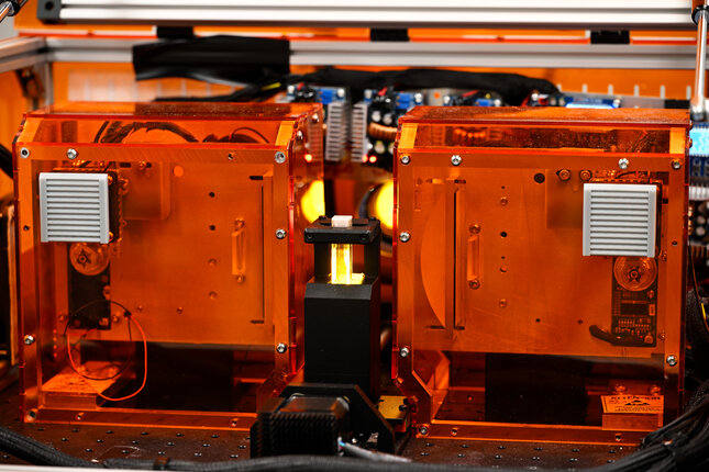
"For now, we view the technology still as a hackerspace." This pioneering spirit is perfectly reflected in the printer, an early tissue printing prototype, whose sheer orange acrylic casing reveals an inside of wires, projectors, copper coils, and tiny digital displays.
While it may seem speculative for now, the detailed and lightning-fast printing of living tissue in a suit-cased-sized, orange 3D printer is completely real. "And our research is a necessary first step for the future of tissue engineering. Right now, it can print physiologically more relevant 3D environments for cell culture, and in the long term, it could help make 3D-printed organs a reality," says Dias Castilho.
Tissue printing with light
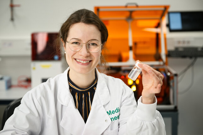
At the heart of the machine sits a tiny cuvette containing a fluid that transforms into a solid as if by magic. But instead of waving a magic wand, Lena Stoecker , who is a PhD of Dias Castilho's brand new Biomaterials Engineering and Biofabrication group, projects beams of light onto liquids to conjure up viable cell-laden geometries.
Stoecker has successfully adapted a novel 3D printing technique called Xolography to print biomaterials. While demonstrating the printer by putting a cuvette with a liquid inside Stoecker explains what drew her to 3D printing tissues: "I first encountered 3D printing as a student assistant during my studies of mechanical engineering and business administration. We employed 3D printing mainly for prototyping and tooling for small series production, and I was fascinated by the technology's possibility to realize (almost) any idea."
I always enjoyed solving the scientific challenges associated with the technologies, but I sometimes missed my personal "higher aim", which I found in turning towards biomedical applications with their clear goal of eventually improving health.
- Lena Stoecker
Biomedical challenges
It is no surprise Stoecker gravitated towards tissue engineering, as it is by nature a multidisciplinary field combining the expertise of molecular biologists, engineers and designers.
The biggest trifold challenge facing tissue engineers everywhere is to create viable 3D tissues that closely resemble the natural environment of cells, to create them fast, and to do it precisely. This is the holy grail. "There was a big hype around 3D printing for biomedical engineering, but technologies failed to meet the high expectations," Stoecker explains. "My dream for Xolography would be to develop into a technology that is actually able to create tissue and organ models to study disease and develop cures."
A technique from the field of design
Xolography is a groundbreaking fusion of engineering, physics and chemistry, where light is used to 3D print liquid polymers. It harnesses the power of intersecting light beams of distinct wavelengths within a light-reactive fluid. As light rays converge, they turn the fluid into a detailed, solid 3D object the size of a gummi bear in under a minute.
The technology was developed by German chemist Stefan Hecht and physicist Martin Regehly, who further adapted it for diverse applications in their spin-off business Xolo. Four years ago, Hecht mused about Xolography potentially being used for generating complex biological structures .
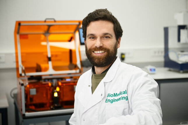
Dias Castilho: "Four years ago Xolo was looking to advance its technology into biomedical applications, while my team was searching for a disruptive technology that could potentially offer high resolution, fast manufacturing speeds, and scalability - so it's a perfect marriage."
Today, the TU/e-researchers at the Biomaterials Engineering and Biofabrication group made printing tissue with light a reality. Hecht and Regehly follow the findings of the research group with interest as they are the first scientists to use this technology to print living materials in the world.
101 printen met licht
Most known light-printing techniques are either slow and precise, or fast but lacking detail. Xolography uses visible light and UV light to 3D print materials fast and incredible detailed, as small as 20 µm, the size of a human cell.
How does this printing technique work? It is different from other 3D printing techniques where you print layer over layer. With Xolography you print by 'curing' (making solid) a liquid. The shape of the resulting solid tissue is controlled by projecting slices of an image of the desired tissue onto a light sheet, much like cinemas use rolls of film to screen movies on the big white screen.
For this to work you need two different types of light, UV light and visible light: The fluid reacts or "switches on" if exposed to UV light. When it is switched on, a series of images of the desired structure is projected onto the liquid with visible light. By polymerization the liquid solidifies into the final shape.
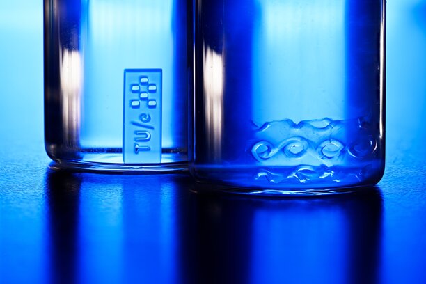
3D samples printed with the Xolography printer: Gyroid (NIPAAm/GelMA hydrogel), TU/e logo with chess pattern (PEGDA hydrogel). Photo: Bart van Overbeeke
That did not happen overnight, as the researchers had to overcome some additional challenges to adapt Xolography to printing living tissue, Dias Castilho mentions a few of them:
"The materials used must be biocompatible, for one. Besides the hydrogels we were developing for the process, we found that the photoinitiator system itself was not very cell-friendly and had to be replaced. In close collaboration with the company, we developed and optimized the material formulations to ensure they are safe for biomedical applications."
Printing scaffolds to grow cells in the lab
Stoecker printed hydrogel scaffolds that can be used as temporary support structures to grow cells in the lab. She says: "To successfully grow tissue, we aim for the hydrogel scaffolds to contain features that mimic the natural environment of, for example, bone marrow cells."
"We were able to print detailed scaffolds with pores in the range of 100 μm-1 mm which could ensure nutrient supply throughout the scaffold during cell culture. Tiny raised elements could be printed down to only 20 μm, in the size range of a human cell."
Tuning 3D material properties with light
Printing on a small scale alone is not enough to mimic natural tissue and exert precise control over the cell's behaviour. "Natural tissues show a variety of properties, for example, they are stiffer in one place and more flexible in another. Existing techniques print objects that are more homogenous," says Stoecker.
"We succeeded in creating materials where we control the properties completely in 3D, so we can create stiffer and flexible regions where we desire them." By varying the projected light intensity, the researchers could tightly control tissue properties.
Artificial muscles
Dias Castilho says: "It has been very hard to replicate materials that can change their shape - and change it back again, which is essential to create tissues that functions like organic tissue:"
The team managed to implement thermally responsive hydrogels to allow the creation of 4D printed structures, where the fourth dimension is time. "These materials can change shape or properties over time in response to temperature changes, enabling more complex and functional tissue constructs," says Dias Castilho, "such as artificial muscles that can flex and extend in response to subtle temperature changes."
Dias Castilho: "We have now demonstrated that this technology could unlock exciting possibilities in healthcare by producing more realistic and functional tissue models and implants."
The results published in Advanced Materials are considered a hallmark publication. "We believe this is a foundational paper," says Dias Castilho. "I believe insights in the paper will help to advance light-based high-resolution fabrication of cell-laden hydrogels with programmable mechanical properties and shape. In the next phase, in vitro models and bioprint solutions for tissue repair will be improved."
Stoecker adds: "I am well aware that our research has to still go a long way to reach the clinic, but I like the idea, that maybe one day, the techniques we develop in the lab will contribute to improve the health and thereby the life of somebody."


