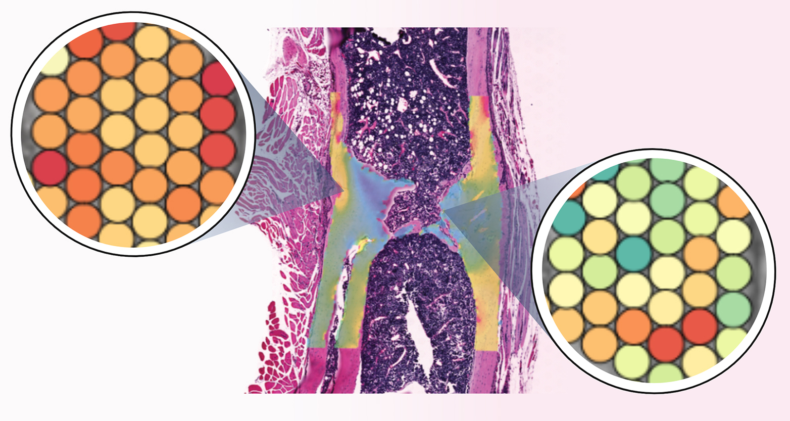
Researchers aim to use vibrations to stimulate bone growth. Now, a new study paves the way for developing new therapies that may one day benefit patients suffering from bone fractures and age-related bone loss.
In brief
- Researchers from ETH Zurich have presented high-resolution spatial data on gene activity in bone cells for the first time.
- By doing so, they have shown that mechanical stimuli influence the activities of genes in bone. Stimuli of this kind could therefore be used in a targeted manner in therapeutic applications.
- The results could pave the way for new therapeutic approaches to combating bone loss and allowing faster bone healing following a fracture.
Bone does not just grow in any which way - rather, the bone cells respond to external forces. If bones are subjected to targeted mechanical loading as they heal following a fracture, they can potentially become larger, denser and more stable than they were before the fracture occurred. This effect was demonstrated in mice three years ago by researchers led by Ralph Müller, a Professor at the Department of Health Sciences and Technology. In that work, the scientists used special plates to fix bone fractures in place, allowing the two parts of the healing bone to be pressed against each other cyclically for a few minutes several times a week in a form of vibration therapy.
However, the mechanisms by which mechanical stimuli influence bone have remained elusive. "Only if we understand these mechanisms can we use them as the basis for developing new therapies," says Neashan Mathavan, a researcher in Müller's group and lead author of a new external page study . Mathavan is talking not only about the healing of bone fractures but also about how fractures can be prevented, particularly in older people. In old age, bone density decreases and bones become more vulnerable to fractures. "Ideally, we need new therapeutic approaches to delaying the breakdown of bone in old age."
Gene activity deciphered point by point
Mathavan, Müller and their colleagues have now carried out a highly detailed investigation of where which genes are active in a healing bone. This work was once again carried out in mice with a broken thigh bone, whose healing was supported with vibration therapy. The researchers determined - to a high level of spatial resolution - which genes are active at each point in the bone, and which are not. They then combined this three-dimensional atlas of gene activity with information on forces acting at the respective locations, which they calculated using computer simulations. "For each point in the bone, we now know what mechanical conditions exist there, where bone is being formed and where bone is being broken down," explains ETH professor Müller.

This approach allowed the researchers to show that certain genes are active specifically in the areas of bone that experience strong mechanical stress. These genes include some that contribute to the formation of the collagenous matrix of bone and some that promote bone mineralisation. Conversely, genes that inhibit bone formation are not active at those locations, but rather in areas that do not experience mechanical stress.
The scientists now plan to use their findings to propose new therapeutic approaches that allow fractures to heal better and bones to remain strong even in old age. In their research in mice, they will now put a special focus on the topic of bone aging.
It may be possible to use drugs in a targeted manner to activate or inhibit the desired genes, but Müller believes vibration therapy or a combination of the two is equally conceivable. "We will see which direction it takes," says the researcher, although he expects vibration therapy to offer certain advantages. "It's likely that vibration therapy will involve fewer side effects than treatment using drugs."
Reference
Mathavan N, Singh A, Correia Marques F, Günther D, Kuhn GA, Wehrle E, Müller R: Spatial transcriptomics in bone mechanomics: Exploring the mechanoregulation of fracture healing in the era of spatial omics. Science Advances, 1 January 2025: doi: external page 10.1126/sciadv.adp8496


