Everything the brain does-from storing memories to interpreting sights to regulating emotions-requires energy, all produced by cellular organelles called mitochondria.
However, surprisingly little is known about the distribution and diversity of the brain's tiny energy processors and how they influence brain health. For instance, how many mitochondria does the brain have? Are they uniformly distributed across the whole brain? Are all brain mitochondria the same? Do changes in the brain's mitochondria affect mood, cognition, and the development of neurological and psychiatric conditions?
To begin answering these and other questions, Columbia University researchers have created MitoBrainMap, the first-ever atlas of the brain's mitochondria.
"There's an emerging notion that energy is really important to health," says study leader Martin Picard, associate professor of behavioral medicine (in psychiatry, neurology, and the Robert N. Butler Columbia Aging Center), who led the study with Michel Thiebaut de Schotten, research director at the University of Bordeaux. "But we don't have a way to look at bioenergetics across the entire human brain."
"MitoBrainMap marks a milestone toward understanding the energy landscape underlying brain health, with implications for tracing the origin of neurodegenerative and neuropsychiatric disorders and developing new approaches to treatment."
Making the atlas
A major barrier to understanding the brain's energy landscape is the "scale gap" between microscopic studies of mitochondria within individual cells and macroscopic neuroimaging studies, such as magnetic resonance imaging (MRI), that look at the entire brain.
To bridge this gap, the researchers obtained a section of a frozen human brain and diced it into 703 3x3x3mm cubes, a size that corresponds to the resolution of neuroimaging scanners.
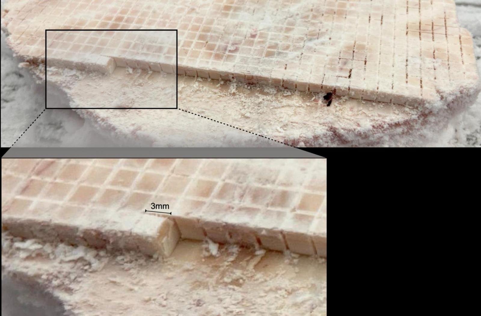
Analysis of mitochondria within 703 3x3x3mm cubes of a human brain created the first energy map of the brain. Image provided by Martin Picard / Columbia University Vagelos College of Physicians and Surgeons.
The team then determined the mitochondrial density and energy transformation capacity within each cube-roughly the size of a large grain of sand-yielding an energy map of the entire slice.
Computational modeling was then employed to extrapolate the findings from the single slice to the whole brain-a necessary compromise since analyzing each one of the brains ~50,000 voxels would have taken many years to accomplish.
What the mapping revealed
The mapping effort revealed that mitochondria differ not only by cell type, but also by the brain region. "These differences are remarkable since all mitochondria originate from the same "mother" pool of mitochondria in the oocyte," says Anna Monzel, a computational research scientist who was involved with the single cell mitochondrial phenotyping ('mitotyping') component of the study. "But during development they specialize in a cell type-dependent manner to subserve bioenergetic demands and guide the acquisition of specific cellular phenotypes."
The researchers also found that newer brain regions, which distinguish humans from other species, not only contained more mitochondria, but these mitochondria were specialized for more efficient energy production, in keeping with the high energetic costs of these regions compared with evolutionarily older regions.
Next steps
Additional research is now needed to test, validate, and apply MitoMapping in various research and clinical contexts.
"If validated, our model could be used to estimate the functional properties of mitochondria in the live brain using standard MRI scans, essentially creating the first non-invasive way of peering into the biology of mitochondrial bioenergetics in the human brain," says the study's first author Eugene Mosharov, research scientist in the Department of Psychiatry.
Such scans would enable scientists to explore relationships between mitochondrial function and brain activity during cognition, development, disease states, and psychological states."Our model could be used to estimate the functional properties of mitochondria in the live brain using standard MRI scans, essentially creating the first non-invasive way of peering into the biology of mitochondrial bioenergetics in the human brain."
MitoBrainMap is just the first version of the brain's energy landscape. The researchers are currently analyzing nine different brain regions in some 500 brains, which will help them determine how patterns of mitochondrial distribution and specialization differ among individuals and improve the map's prediction accuracy.
"Energy is the missing dimension of biomedicine," Picard says. "If you think of health as energy, it inspires you ask different questions. How much energy does it cost to heal the brain? Does the food you eat influence your mitochondria? Do energy constraints affect normal brain function, or the development of Alzheimer's disease? This MitoBrainMap v1.0 is a step toward understanding the energetics of the brain and the experiences it allows us to have."
References
Additional information
"A human brain map of mitochondrial respiratory capacity and diversity," was published March 26 in Nature.
All authors: Eugene V. Mosharov (Columbia), Ayelet M. Rosenberg (Columbia), Anna S. Monzel (Columbia), Corey A. Osto (UCLA), Linsey Stiles (UCLA), Gorazd B. Rosoklija (Columbia), Andrew J. Dwork (Columbia), Snehal Bindra (Columbia), Alex Junker (Columbia), Ya Zhang (Columbia), Masashi Fujita (Columbia), Madeline B. Mariani (Columbia), Mihran Bakalian (Columbia), David Sulzer (Columbia), Philip L. De Jager (Columbia), Vilas Menon (Columbia), Orian S. Shirihai (UCLA), J. John Mann (Columbia), Mark Underwood (Columbia), Maura Boldrini (Columbia), Michel Thiebaut de Schotten (University of Bordeaux), and Martin Picard (Columbia).
This project was supported by the NIH (grants RF1AG076821 and RF1AG057473); the Baszucki Brain Research Fund; Columbia University Department of Psychiatry; the JPB Foundation; HORIZON- INFRA-2022 SERV (Grant No. 101147319) "EBRAINS 2.0: A Research Infrastructure to Advance Neuroscience and Brain Health," by the European Union's Horizon 2020 research and innovation programme under the European Research Council (ERC) Consolidator grant agreement No. 818521 (DISCONNECTOME); the University of Bordeaux's IdEx 'Investments for the Future' program RRI 'IMPACT'; and the IHU 'Precision & Global Vascular Brain Health Institute, funded by the France 2030 initiative (ANR-23-IAHU-0001).


