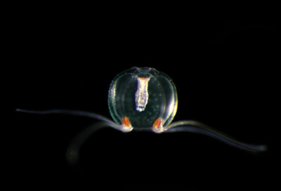
 Figure 1: RIKEN researchers have derived a photostable and bright green fluorescent protein from the Japanese jellyfish Cytaeis uchidae. © 2022 Kuroshio Biological Research Foundation
Fluorescence imaging of biological samples stands to benefit greatly by a RIKEN discovery of a fluorescent protein derived from a Japanese jellyfish that maintains its brightness even when illuminated by strong light1.
Proteins that give off green light when illuminated are powerful tools for imaging fine structures within living cells. Researchers can attach such fluorescent proteins to target structures they are interested in, which then light up when blue light is shone on them.
However, researchers find themselves in a bind-they want to use as little fluorescent protein as possible so that it doesn't interfere with normal cellular processes, but that necessitates using strong illumination in order to obtain high-quality images. The trouble is that when strong light is shone on a fluorescent protein, its brightness drops off rapidly due to a process known as photobleaching. To complicate matters, there is a trade-off relationship between brightness and photostability: increasing one will almost inevitably reduce the other.
Now, Atsushi Miyawaki of the RIKEN Center for Brain Science and his co-workers have discovered a fluorescent protein that flouts this trade-off relationship: it offers both high brightness while being roughly ten times more photostable than the best commercial fluorescent proteins.
Appropriately named StayGold, the fluorescent protein is derived from a naturally occurring fluorescent protein found in Cytaeis uchidae, a tiny jellyfish found off the coast of Japan (Fig. 1).
There was an element of serendipity in the discovery. "We noticed that the fluorescent protein from the jellyfish was photostable but very dim. And I wasn't optimistic about making the protein brighter while keeping that photostability, because I simply believed the tradeoff," recalls Miyawaki. "However, to our surprise, we were able to increase both the protein's photostability and its brightness. So could have our cake and eat it too."
The team demonstrated the usefulness of StayGold by using it to image the endoplasmic reticulum network and mitochondria in cells with enhanced spatiotemporal resolution and length of observation. They also used it to image the spike protein of SARS-CoV-2, the virus that causes COVID-19, in infected cells.
The intense interest generated by the study is reflected in the fact that it has been accessed more than 44,000 times since publication in late April. Researchers wanting to try the protein can obtain it from the RIKEN BioResource Research Center (http://en.brc.riken.jp).
Since it remains unclear why StayGold can both be bright and stay bright under illumination, Miyawaki and his team intend to investigate the mechanism behind this.


