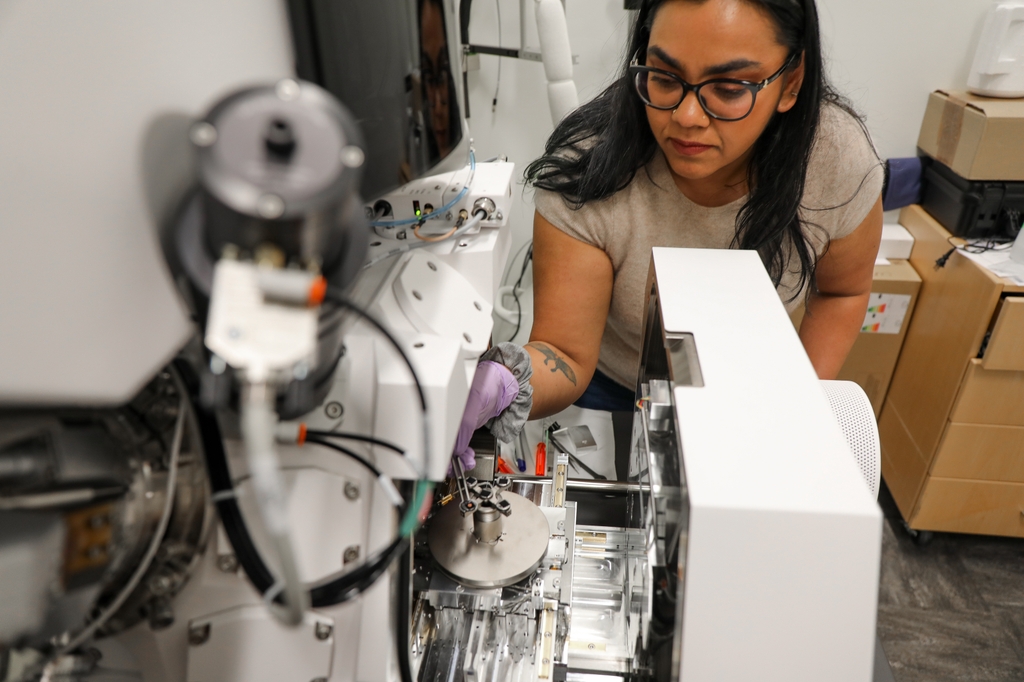
When Claudia López, Ph.D., first arrived at Oregon Health & Science University in 2003 as a postdoctoral fellow, she never imagined the trajectory her career would take. Originally trained in biology and biophysics, Claudia's curiosity about microscopy technology and her deep commitment to research would lead her to pivotal leadership roles in two of OHSU's most groundbreaking initiatives: the Pacific Northwest Center for Cryo-EM and the Multiscale Microscopy Core.
"I came to OHSU for a short-term postdoc and ended up staying," she said. "My journey started with electron microscopy and it's been a fascinating path of learning and growth ever since."
Today, López leads the OHSU Multiscale Microscopy Core and is co-director of the Pacific Northwest Cryo-EM Center, which together form an unmatched resource for researchers studying everything from molecular biology to cancer and infectious diseases. Both centers are located in OHSU's Robertson Life Science Building, which has a specialized basement with a 5,000 square-foot concrete pad, supported by more than 70 pylons, each 200 feet long, to hold the heavy and sensitive microscopes.
In January, the team will launch a new microscopy suite for the Multiscale Microscopy core that will expand by an additional 1,600 square feet the space OHSU is dedicating to high-end microscopy.
Building the local facility
In 2013, OHSU established the Multiscale Microscopy Core, an initiative sparked by a vision from Joe Gray, Ph.D., professor emeritus of biomedical engineering in the OHSU School of Medicine, who recognized the need for a diverse set of imaging tools that could span cellular, molecular and even atomic-level analysis. López was there at the beginning, managing a small but growing facility with just a couple of electron microscopes.


