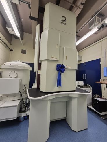
The University of Oxford's Department of Materials celebrated a new chapter in its microscopy facilities with the arrival of a bespoke £3 million Transmission Electron Microscope. The JEOL GrandARM300F instrument will support cutting-edge research across the University's Departments and Divisions, besides teaching the next generation of microscopists. The instrument was officially launched during the 100 birthday celebrations of Professor Peter Hirsch, one of the University's foremost materials scientists.
Housed in the David Cockayne Centre for Electron Microscopy , the 3.5-metre high microscope will support the facility's purpose of helping researchers obtain the highest possible imaging data of samples. Using beams of electrons accelerated to up to 300 kV, the new GrandARM300F microscope is capable of magnifying samples up to 50 million times, enabling individual atoms to be visualised.
 The GrandARM300F Transmission Electron Microscope. Credit: Caroline Wood.
The microscope was officially opened by Vice-Chancellor Professor Irene Tracey on Monday 17 March 2025 during a special event to celebrate the 100th birthday of Sir Peter Hirsch , Emeritus Professor of Metallurgy who was Head of Oxford's Department of Materials from 1966 to 1992.
A key attribute of the GrandARM300F is its versatility, being able to rapidly switch between different modes (including Transmission Electron Microscopy, Scanning Transmission Electron Microscopy and X-ray spectroscopy). This allows researchers to capture different forms of information on the same sample in real-time.
The GrandARM300F has also been specially designed to analyse 'beam-sensitive' samples that are highly susceptible to degradation. Features include a cryogenic mode that can cool samples down to the temperature of liquid nitrogen (around -190°C) and highly precise electron beam control, allowing it to be reduced to as little as 60 kV.
Dr Neil Young, Senior Electron Microscope Facility Manager for the David Cockayne Centre for Electron Microscopy, said: 'The new microscope is designed to enable users to quickly switch between modes and techniques. This provides multi-modal datasets, elevating the characterisation of materials to new levels. Furthermore, the microscope is optimised for low-dose and low-voltage studies, allowing us to analyse sensitive samples which would otherwise be damaged by the electron beam.'
The GrandARM300F will support University research across a range of strategic areas, including:
- Energy materials for the Net Zero transition,
- Drug delivery systems, for instance in chemotherapy,
- Disease mechanisms in biological tissues,
- Carbon capture,
- Elemental analysis,
- Semiconductor research,
- Polymer research and metal-organic frameworks.
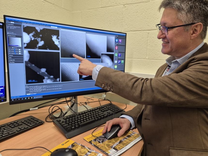
 Dr Gerardo Martinez points to individual atoms imaged using the new GrandARM300F Transmission Electron Microscope. Image credit: Caroline Wood.
'Besides high-end research, this new microscope will also play a vital role in teaching and training the microscopists of the future' said Dr Gerardo Martinez, the Transmission Electron Microscopy Support Scientist for the David Cockayne Centre for Electron Microscopy. 'There are few other places in the world where students have the opportunity to operate a TEM microscope of this calibre themselves, giving an immensely valuable learning experience.'
 Professor Sir Peter Hirsch. Credit: University of Oxford/Richard Lofthouse.
As with the other instruments within the David Cockayne Centre, the GrandARM300F is also available for external researchers to use (for information, see the Centre's website ).
The new instrument builds on the Department's strong track record in materials research, including advanced techniques and imaging methods. It was Professor Sir Peter Hirsch who commissioned the first JEOL electron microscope for the department fifty years ago. He is renowned for his pioneering work in Transmission Electron Microscopy and its application to the study of materials, especially metals and alloys. In particular, he and his group developed the theory of electron diffraction contrast imaging, which is used to monitor defects introduced during materials processing. This gives insight into mechanical properties and is critical for the development of new materials including semiconductors, nuclear radiation-resistant alloys, and high-temperature alloys for jet engines. Professor Hirsh was knighted in 1975. Between 1988 and 1996 he was founder Chairman of Isis Innovation, the technology transfer company of the University of Oxford.


