We're surrounded by an unseen atomic world that governs life and the technologies we use every day, such as batteries and computers.
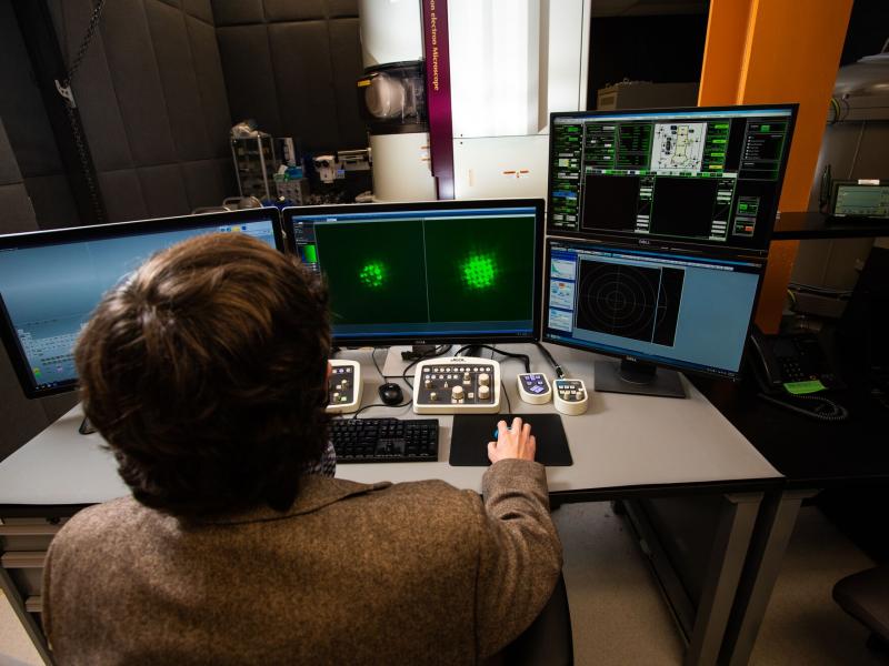
Scientists have developed powerful microscopes that use electron beams to peer into this world, revealing how tiny atomic motions and defects have a big impact on materials and chemical processes. Still, many questions remain out of reach of today's microscopes, requiring us to reimagine the tools used to survey the atomic landscape.
An international research team led by Pacific Northwest National Laboratory (PNNL) has published a vision for electron microscopy infused with the latest advances in data science and artificial intelligence. Writing a commentary in Nature Materials, the team proposes a highly integrated, autonomous, and data-driven microscopy architecture to address challenges in energy storage, quantum information science, and materials design. They describe how this approach can provide new insight into materials properties, permitting experiments at a vast scale not possible today.
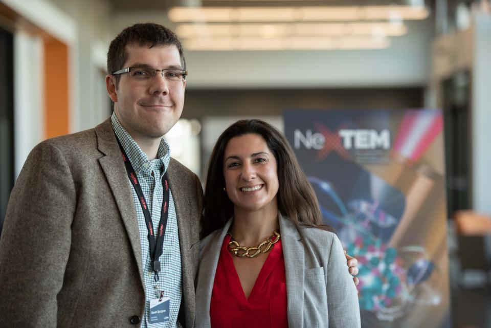
"Materials science and microscopy are undergoing a radical transformation," explains Steven Spurgeon, a PNNL materials scientist and lead author on the publication. "Advanced data science techniques are starting to reshape how experiments are planned, conducted, and interpreted, enabling us to extract deep, truly statistical information about very complex processes for the first time."
Microscopy limitations and information overload

Electron microscopy has long played a pivotal role in materials characterization down to the level of single atoms, aided by developments such as improved electromagnetic lenses. However, the field as a whole, Spurgeon says, has not yet adopted data science methods that have revolutionized other domains, such as single particle cryo analysis and X-ray crystallography.
Traditional microscopy is constrained in its ability to effectively analyze many representative samples and integrate large volumes of multidimensional data from high-speed detectors. Another limitation is the difficulty in accessing data from past experiments, which keeps the data from being directly incorporated in the experiment design and analysis process.
"We are rapidly approaching the point of data saturation," says Mitra Taheri, co-author of the publication and director of the Materials Characterization and Processing Laboratory at Johns Hopkins University, with an appointment at PNNL.
"To fully harness these unprecedented volumes of data we need to completely rethink how experimentation is conducted in microscopy," Taheri says. "Not only do artificial intelligence and machine learning tools allow us to manage data flow, but also they enable more innovative microscopy solutions going forward."
Making microscopy data internationally accessible

In their article, the research team proposes to integrate artificial intelligence and machine learning into each step of the microscopy workflow. They argue strongly for the development of a new microscopy infrastructure-advanced via multi-institute national technology initiatives-that makes data more accessible to research organizations worldwide. In this way, past measurements could be used to select techniques and interpret results on the fly, informing autonomous decision-making algorithms. Large libraries of past experiments could be used to highlight latent features and offer guidance for the user. A strength of this "crowdsourced" approach is that it is highly scalable, less prone to operator bias, and more repeatable, which will translate into improved results.
Ultimately, the team hopes to show the immense, untapped potential of data science and its critical role in unlocking the full power of electron microscopy.
"As a community we have to rethink the way microscopy is done at every level, from the domain expert to the instrument designer, if we want to solve the challenges of the next decade," Spurgeon says. His work at PNNL includes leading a Nuclear Process Science Initiative project on high-resolution studies of radiation damage in oxides, which supported this work.
The commentary, published in the October 26, 2020, online edition of Nature Materials (DOI: 10.1038/s41563-020-00833-z), is the result of the first Next-Generation Transmission Electron Microscopy (NexTEM) workshop held at PNNL in 2018.
The team of authors includes Colin Ophus and Haimei Zheng, Lawrence Berkeley National Laboratory; Lewys Jones, Trinity College Dublin, Ireland; Amanda Petford-Long, Argonne National Laboratory; Sergei V. Kalinin, Oak Ridge National Laboratory; Rafal E. Dunin-Borkowski, Forschungszentrum Jülich, Germany; Norman Salmon, Hummingbird Scientific; Khalid Hattar, Sandia National Laboratories; Wei-Chang D. Yang and Renu Sharma, National Institute of Standards and Technology-Gaithersburg; Ann Chiaramonti, National Institute of Standards and Technology-Boulder; R. Lee Penn, University of Minnesota; Mitsuhiro Murayama, Virginia Tech; Mitra Taheri, Johns Hopkins University; and Matthew J. Olszta, Dongsheng Li, Xin Zhang, Yingge Du, Libor Kovarik, Edgar Buck, and Steven Spurgeon, PNNL.


