The development of maternal egg cells is pivotal for survival - but also precarious. During meiosis, the DNA-containing chromosomes can easily be broken or lost, causing infertility, miscarriage or genetic disorders like Down syndrome. Scientists have struggled to study these crucial cellular events in humans and other mammals.
"The ovaries are opaque, you cannot see inside them," said Francis McNally, a professor of molecular and cellular biology at the University of California, Davis.
Scientists usually have to study cells outside the body, and hope that they're seeing the normal, natural process. But McNally is taking a different approach. He is eavesdropping on egg development as it unfolds inside the mother - mother worms, in this case.
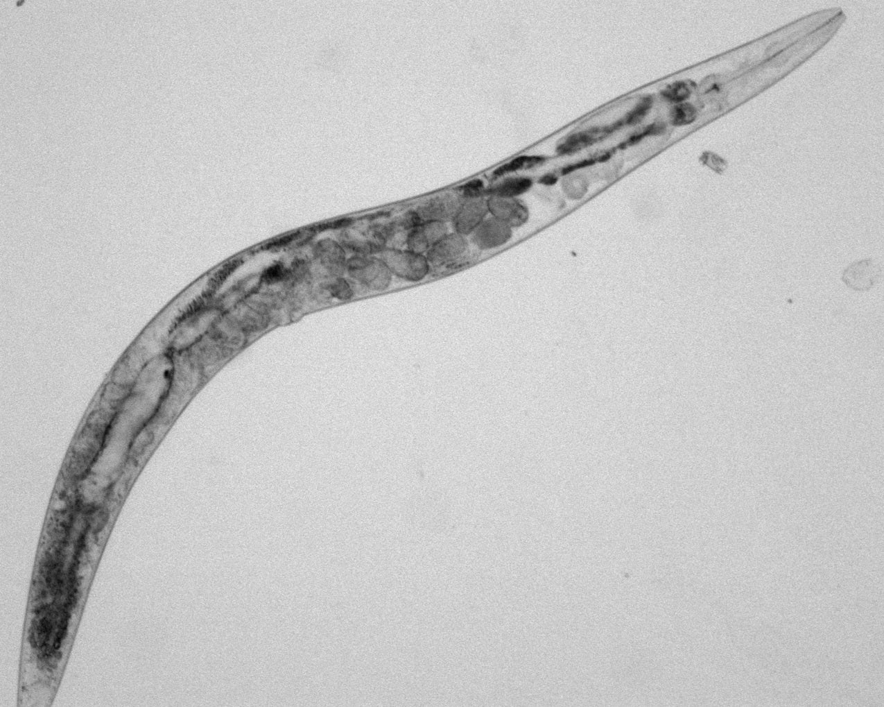
Caenorhabditis elegans is a tiny worm that lives in soil. It is a millimeter long and as wide as several human hairs. It is also transparent.

"It gives us a rare window into something we couldn't otherwise see," McNally said. "You can anesthetize the worm, watch through a microscope, and film the entire process in 45 minutes" as an egg cell develops and is fertilized by a sperm.
McNally's study of worms could improve our understanding of human reproduction and how fertility problems might be diagnosed, prevented or treated.
A distant relative is surprising similar
At first glance, C. elegans isn't the place you would expect scientists to look for insights into human biology. Its body consists of just 959 cells. It has no lungs, liver, kidneys, eyes or circulatory system. The cells that turn into sperm and eggs make up more than half its body.
"It's basically a gut and two gonads," McNally said.
But the little worm also shares some surprising affinities with us. Its DNA holds more than 19,000 genes - only a few hundred shy of the lofty human genome. Many of those genes are "homologs," closely related to genes that humans use to guide cell division, egg cell production and other critical facets of life.
McNally studies one of these critical events, called meiosis, which happens as a maternal egg cell develops. The cell starts with a quadruple set of DNA - four of each chromosome. Twice in a row, it has to line up those chromosomes and divide them in half, discarding the ones that aren't going to be kept. The egg cell ends up with only one of each chromosome - in anticipation of the sperm cell that will arrive and deliver a complementary set.
It's a perilous process.
As chromosomes line up in the middle, a mechanized protein scaffold, called the spindle, assembles on either side of that line. On each side, it resembles a squid with its tentacles stretched out - their tips attached to one half of the chromosomes. At some point, the tentacles shorten and the matching chromosomes are drawn apart.
Those tentacles, called microtubules, assemble from thousands of tiny protein segments. Their growth, shortening and attachment to chromosomes is choreographed by dozens of other proteins, each too small to see under a microscope.
Delicate dance of chromosomes
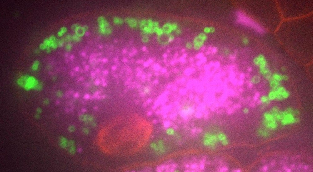
McNally is using worms to tease this process apart. He keeps dozens of different worm strains, each with genes modified to make a particular protein that is attached to a jellyfish protein, causing it to glow a specific color when exposed to ultraviolet light.
McNally puts the transparent worm under a microscope, focuses on a single egg cell and records videos as the different proteins - marked by their red, green or blue glow - delicately maneuver about. By knocking out one or a few genes at a time, and watching what goes wrong, he can deduce the role of each protein.

Over the years, he has repeatedly examined a protein called katanin (named after the traditional Japanese sword, katana), which cuts the microtubule "tentacles." In 2014, he found that katanin must constantly prune the tentacles - otherwise they grow haphazardly like the arms of a kraken, pushing the chromosomes out of position.
Katanin plays other roles. When a sperm fertilizes an egg, the maternal cell has not yet finished meiosis. It still has two of each chromosome and must separate those chromosome pairs and discard the unused set - all while the sperm DNA sits and waits inside the cell.
This early arrival of the sperm happens in most animals, from worms to mice to humans.
If the sperm and egg DNA mingle too soon, then some of sperm's chromosomes could end up being discarded along with the extra female ones - dooming the offspring.
In a paper published this July, he found that katanin and two other proteins, kinesin-13 and ataxin-2, prevent this from happening.
And in August, McNally reported another surprise finding, unrelated to katanin. People have speculated about what triggers the microtubule tentacles to assemble on cue. A number of proteins seem to play a role. But McNally found that microtubule growth is also driven by another, unseen force.
Those protein segments assemble inside a small transparent sack, which crowds them together. That crowding actually triggers them to stick together and assemble on their own.
When McNally makes these discoveries in worms, it gives other researchers a chance to see if the same things are happening in mammals.
"We are looking for things that people are not studying in humans and mice," he said.
Not every discovery will provide new insights into human fertility. But they all reveal the diversity of evolution for solving some of life's hardest challenges.
"Some things that you find in worms will be exactly the same in humans, and some will be different," said McNally. "Both are important and interesting."


