At the Oregon Health & Science University Advanced Imaging Research Center, or AIRC, scientists who specialize in magnetic resonance imaging conduct their research and serve as advisers and collaborators to other researchers who use MRI techniques.
Within the Lamfrom Biomedical Research Building on OHSU's Marquam Hill Campus, the AIRC features equipment found in few places around the world. This includes a 12 Tesla MRI for studying animal models and a 7 Tesla MRI for studying human subjects, equipment that allows for faster and more detailed images.
This advanced imaging technology makes OHSU a critical resource for scientists conducting basic, clinical and translational research on development across the lifespan and diseases and conditions such as brain cancer, strokes, multiple sclerosis, dementia and reproductive issues.
"Imaging at the AIRC was conceived from the beginning as a hybrid construct, really unique, because it is a mix between an academic department and a shared resource," said William Rooney, Ph.D., AIRC director and a professor of behavioral neuroscience in the OHSU School of Medicine.
"We have these big instruments, and our faculty are here to develop them, maintain them, do their own research, but they are also available to the broader research community, which our faculty also help facilitate."
Expanding to meet a growing need
In the late 1990s, as the use of MRIs in clinical practice became increasingly common, the need for increased access to imaging technology became clear to the OHSU neuroscience community. The few MRIs at OHSU at the time were needed for patient care. A group of OHSU faculty members developed a plan to create a research-dedicated MRI center which began to crystalize after receiving funding from the Office of National Drug Control Policy for the purchase of an MRI instrument. This, together with accessing Oregon Opportunity tobacco settlement funds, enabled the creation of the Advanced Imaging Research Center.
In 2003, Charles Springer, Ph.D., was recruited to serve as the director of the newly formed center. He recruited many of the researchers who are still with the center, including Rooney. Work began to write grant applications and secure funds, including a W. M. Keck Foundation award, to obtain the imaging equipment and eventually build the Lamfrom Biomedical Research Building, as well as the West Campus satellite AIRC facilities for the Oregon National Primate Research Center. In Lamfrom, the AIRC now occupies approximately 9,500 feet, including space for a 3 Tesla MRI suite, 7 Tesla whole-body MRI and 12 Tesla MRI instruments, with their associated control rooms and electronics areas. There are also faculty and staff offices and additional office spaces for postdoctoral associates and graduate students. There is a conference room, a chemistry laboratory, and much larger spaces for the electronic shop and image processing laboratory. There is also a mock scanner for fMRI training and control studies.
"OHSU went from an institution that didn't have any dedicated research MRI to one of the most comprehensive imaging centers in the world," Rooney said. "It was a bold move for the institution."
Rooney said it would not have happened without the leadership of then-Senior Vice President of Research Daniel Dorsa, Ph.D., and many faculty members who recognized that this was the missing link to conduct the kind of research needed to make significant advances in human health.
"OHSU faculty were very competitive," Rooney said. "They were getting grants and really one of the only things holding them back was just access to those dedicated resources for research. So, they made it happen."
Here are some highlights from a few AIRC researchers using OHSU imaging to conduct important research:
Charles Springer, Ph.D.
Springer brings more than 35 years of experience in in vivo magnetic resonance scientific research to the AIRC. Before being recruited to be the founding director of the AIRC, he was a chemistry professor at Stony Brook University and founding director of the High-Field MRI Laboratory, Brookhaven National Laboratory Chemistry Department.

He helped build the AIRC in the early 2000s, recruiting faculty, including current director Rooney, and helping select the imaging equipment such as the 3, 7 and 12 Tesla MRIs, which allow for faster, more detailed scans and higher quality images.
In 2009, Springer stepped down as director because he wanted to focus on research. He is now an adjunct professor of chemical physiology and biochemistry, and biomedical engineering. He had NIH-funding for more than 30 years. His current passion is an innovation developed at OHSU called metabolic activity diffusion imaging, or MADI. MADI uses MRI to scan fine, metabolic details in the human brain and other organs. This discovery opens new possibilities for detecting cancers and revealing if a tumor is responding to treatment. In recent publications, Springer and colleagues compared MADI with positron emission tomography, or PET, on subjects with brain tumors. PET uses injected radioactive agents to create images of cellular glucose uptake rates and is a standard for imaging tumor detection.
In studies with principal investigator Martin Pike, Ph.D., an associate professor at the AIRC, the team showed in a rat brain tumor model that MADI, which does not require injecting an agent, diagnosed brain tumors in rats with high accuracy.
MADI is noninvasive, and like PET can measure metabolic activity but can do so with higher spatial resolution images.
"If you're doing therapy, especially investigating for new drugs, you really want to know the metabolism, that is how you decide to target new drugs," he said. "MADI's ability to measure metabolic activity should guide development of new therapies."
While the team has tested MADI in smaller human trials, Springer said much larger clinical trials are needed before the method becomes part of standard of care. OHSU has obtained a patent for MADI.
Martin Pike, Ph.D.
Pike's research focus is on the pathophysiology of brain diseases, including malignant glioma and stroke, using MRI/MRS in tandem with other approaches. He joined OHSU as a visiting scholar at the Advanced Imaging Research Center in 2007 and became an associate professor at the OHSU Knight Cancer Institute in 2008.

His lab has employed dynamic contrast MRI approaches to investigate anti-angiogenic/anti-tumor treatment strategies employing mouse models of malignant glioma, a cancerous tumor that develops in the brain from glial cells. Pike uses the 12 Tesla MRI, an instrument that has a magnetic field 120,000 times stronger than that of the Earth. It is used for human health studies in small animal models. Like most of the researchers at the AIRC, Pike splits his time between his own work and assisting other researchers throughout OHSU with their projects.
"We've always had an edge on imaging animals because of the high field MRI," Pike said. "We recently funded an upgrade that included a special detector probe called the Cryoprobe, which very few facilities have."
This detector probe is cooled to cryogenic temperatures, which gets rid of much of the thermal noise and results in a three-fold increase in MRI signal to noise. What this means is a much higher resolution in a small animal model, such as a mouse or rat.
"There are structures we couldn't see very well before," Pike said. "So, this has opened up quite a few new projects, particularly with those investigators who are interested in brain white matter structures" (White matter consists of the connections that allow nerve cells in the brain to communicate with each other.)
"With improved resolution, researchers can now more effectively employ imaging strategies to track pathology in diseases such as stroke, multiple sclerosis and dementia," he said. "There is a pathology associated with aging, stroke and dementia called cerebral microbleeds, which are also very tiny in mice, and these now can be effectively observed and quantified."
His own research currently is focused on using Springer's MADI invention to measure metabolic activity in the brain.
"We can pull out cell size and density with MADI, but we can also measure the rate of water movement in and out of cells," Pike said. "This water movement is mostly linked to the movement of sodium and other ions across the cell membrane, and it uses much of the cell's energy. So, what we are measuring is really the cell's energy use."
Measuring this energy-dependent metabolic activity is important because diseases like cancer change how cells produce and use energy. By using a noninvasive imaging method to measure it, Pike and his team believe it could become a valuable tool for diagnosing these diseases in the future.
"We hope to test this not only in cancer, but in stroke and Alzheimer's to see if the cellular metabolic activity is altered and to provide improved ways to diagnose these diseases," Pike said.
Christopher Kroenke, Ph.D.
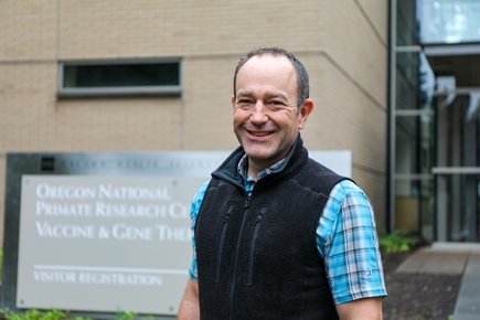
Kroenke is a professor at the ONPRC as well as the AIRC. Like his colleague Pike, Kroenke's laboratory works with the 12 Tesla MRI on animal models. Kroenke's job also involves overseeing the AIRC's nonhuman primate imaging facility at the ONPRC. He often collaborates with primate center colleagues ranging from the research of Victoria Roberts Ph.D., on placenta development, to the work of Verginia Cuzon Carlson, Ph.D., on brain function and connectivity mapping using electrophysiology.

"We have the infrastructure at the ONPRC for translational research so that the imaging methods we develop here can immediately translate to MRI performed with humans," Kroenke said.
His research focuses on brain development. He uses imaging to detect abnormalities in the context of neurodevelopmental science.
"There is epidemiological evidence that the earlier you identify any sort of problem related to brain development, the better the therapeutic intervention is," he said. "This is true even for fetal alcohol spectrum disorders, where there is no known cure. The data show that being able to start any therapeutic interventions as early as possible helps with long-term adverse life outcomes."
In Kroenke's lab, researchers are using fetal MRI techniques to provide high resolution 3D images of the developing brain throughout gestation.
His team uses this method in neuroimaging studies across all ages to help understand abnormal findings in MRI scans of humans. For example, they have found out how alcohol exposure during pregnancy can impact the developing brain and linked their MRI findings to more detailed studies that look at brain tissue and electrophysiology.
His lab is currently using functional MRI to characterize changes in brain function before and after chronic alcohol drinking in nonhuman primates. With others at the ONPRC, his group is integrating new methods, such as DREADDS, or Designer Receptors Activated Only by Designer Drugs, to directly manipulate brain circuitry, and detect the effects with MRI.
Another current project looks at how the folds in the human brain develop. They use animal models to study this process and how problems in the structure and formation of these folds can affect brain function in disorders such as schizophrenia.
"This folding abnormality happens halfway through gestation in a developing fetus," Kroenke said.
And here's the rub: No one knows why the brain folds differently in people with certain disorders compared to a developmentally normal brain.
"It's well documented that the brain folds are different in people with schizophrenia, autism and other neurodevelopmental disorders," Kroenke said. "Using imaging, we can see this folding if we direct our focus on the appropriate developmental stage, which can be as early as during gestation."
His current studies are trying to uncover why and how this folding occurs. Once the researchers have pinpointed that more accurately, earlier interventions could be developed.
"This is an important developmental period, and it's a large step forward because right now, nobody knows how to control it or what influences how these folds form," he said. "Data shows, for example, there is a two-to-three-fold increased risk of being diagnosed with schizophrenia with a specific pattern of brain folds. But we need to understand the why and how so others can develop therapies."
Matthias Schabel, Ph.D.
Schabel, an assistant professor at the AIRC, did not follow the usual path to human health research. By training, he is a physicist, who earned his doctorate from Stanford University in applied physics in the late 1990s. He used his knowledge to study climate data at Remote Sensing Systems in Santa Rosa, Calif. A move to Utah with his wife, a surgeon who completed her residency in Salt Lake City and now also works at OHSU, prompted a career change. Finding no opportunities to continue his climate research in Utah, Schabel jumped from satellite imaging to magnetic resonance imaging in breast cancer research.

"There was a significant gap at that time between qualitative and quantitative MRI research," he said. "So, for example, instead of saying, 'tumors tend to have elevated blood flow,' we were working to get precise values of blood flow."
After Schabel moved to OHSU in 2010, he was contacted by Kroenke to analyze some data from images of placental development in nonhuman primates. It was then he became hooked – here was an understudied area of human development ripe for his mathematical and physicist-based training.
"It sparked my interest," Schabel said. "The placenta is a transient organ that is extremely interesting from several levels. It's a critical organ and it's important that it develops correctly. From the perspective of neonatal well-being, we now know that many problems in pregnancy are associated with placental disfunction of some sort."
Placental dysfunction became a focus of Schabel's research, to see how he could apply his expertise in development of quantitative methods to this organ.
"Historically, the way that people did imaging in the placenta was using existing tools, without fine-tuning them to consider the unique properties of this specific organ," he said. "Scientists had applied tools from MRI that were developed for other areas, like the brain, to the placenta. But the placenta is completely unique. So, we created new methods for imaging and modeling, and ultimately were awarded a patent for some of these techniques."
Key to Schabel's research is understanding how the placenta functions. Oxygen and blood flows to and from the placenta are critical to assessing at-risk pregnancies.
Along with Antonio Frias, M.D., a professor of obstetrics and gynecology in the OHSU School of Medicine, Schabel was part of the Human Placenta Project, a large, multi-year series of studies funded by the National Institute of Child Health and Development of the National Institutes Health. Their study on using blood oxygen level-dependent MRI as a non-invasive technique that can measure oxygen levels in the placenta showed promise for safer ways to detect issues in a pregnancy earlier.
"Beyond the placenta itself not being well-studied, female reproductive health in general has been given short shrift historically," Schabel said, pointing to a condition like preeclampsia, a hypertensive disorder of pregnancy that occurs in about 7% of pregnancies. Despite the seriousness of the disorder, it remains difficult to diagnose and treat, in part because it often shows up after 20 weeks of pregnancy.
"It's not well-understood and therapeutic options are pretty limited," Schabel said. "We now can identify problematic placental function early in the pregnancy. So, the hope is our imaging technologies might be applied to assessing the efficacy of therapeutics as they are developed. Ultimately, the end goal is to mitigate or correct problems with placental development, to track it and correct problems in real time."
William Rooney, Ph.D.
Rooney is the director of the AIRC, leading the center's day-to-day work, and continues his own research.

In the lab, he and his group are studying brain blood vessels by using special MRI scans combined with advanced computational modeling techniques provided by Andreas Linninger, Ph.D., a colleague in Chicago. The combination of MRI with advanced computing is expected to support more detailed assessments of neurodegenerative conditions like multiple sclerosis and Alzheimer's disease. Rooney also looks at how cells use energy and how that relates to tissue damage. He uses MRI methods developed in his lab to explore how problems with energy metabolism can lead to tissue loss.
One specific project that Rooney and colleagues have been a part of is related to muscle composition in muscular dystrophy, a genetic disease that causes progressive muscle weakness and degeneration. The disease is relentless, and much of Rooney's work is focused on using imaging to better understand disease progression.
"One of my guiding principles is, imaging doesn't cure a disease," Rooney said. "But the right imaging approach can be powerful in helping us to better track disease, and in clinical trials to more quickly identify treatments that work."
Rooney and colleagues have used AIRC technology to evaluate genetic therapies in development for muscular dystrophy treatments. Rooney, in collaboration with scientists in Florida, are working to establish MRI biomarkers sensitive for tracking disease progression across all disease stages in Duchenne muscular dystrophy.
Using imaging to establish biomarkers for future treatments is one of the many reasons Rooney is passionate about the work of the AIRC.
"Ultimately, with our imaging, you will be able to show, for example, a metabolic deficit long before any clinical features of the disease are present," Rooney said. "The research at AIRC helps find those biomarkers, which then helps research identify effective treatments much earlier when the disease is easier to reverse. That is ultimately our goal."
All research involving animal subjects at OHSU must be reviewed and approved by the university's Institutional Animal Care and Use Committee (IACUC). The IACUC's priority is to ensure the health and safety of animal research subjects. The IACUC also reviews procedures to ensure the health and safety of the people who work with the animals. No live animal work may be conducted at OHSU without IACUC approval.


