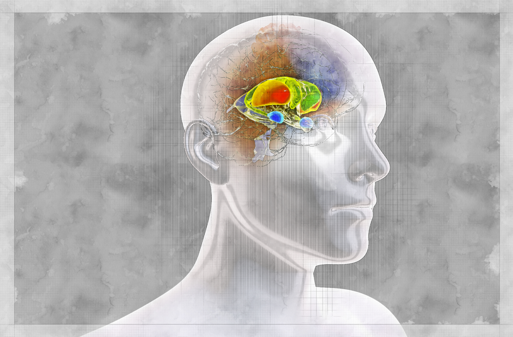
PHILADELPHIA - The neural circuits that form habits in the brain are altered in patients with binge eating disorder (BED), compared to those of patients without such disorders, according to new research led by the Perelman School of Medicine at the University of Pennsylvania. The findings were published recently in Science Translational Medicine.
"This research is the first step in illustrating how complicated conditions like binge eating are-these patients' brains are wired differently," said author Casey Halpern, MD, an associate professor of Neurosurgery and Chief of Stereotactic and Functional Neurosurgery at Penn Medicine and the Corporal Michael J. Crescenz Veterans Affairs Medical Center. "Telling someone with binge eating disorder to not eat so much would be like telling a patient with epilepsy to simply stop having seizures."
Previous research from Halpern suggests that the nucleus accumbens, a brain region involved in processing pleasure and reward, and has been implicated in impulsive behaviors, can be stimulated in patients with BED to disrupt craving-related signals and reduce binging.
"Many patients describe binge eating in response to external food-related cues, like being at a buffet, or internal emotional states, like feeling sad, lonely, or stressed in such a way that feels automatic, like a habit," said senior author, Cara Bohon, PhD, a clinical associate professor of Psychiatry and Behavioral Sciences at Stanford. "So we sought to uncover the role of habit in the development and expression of psychiatric disorders that involve recurrent binge eating, like BED and bulimia nervosa. Our findings may also have implications for other automatic or compulsive behaviors in obsessive compulsive disorder (OCD) and addiction."
Based on previous research into the habit circuits in the brains of small animals, researchers identified the dorsal striatum, part of the brain's reward system, as a location that might play a role in binge eating behavior in humans.
Using functional MRI, researchers evaluated the neural circuitry of habit formation in 34 patients with recurrent binge eating, and compared that with imaging from 19 healthy controls. Researchers measured resting-state functional connectivity (RSFC) between the two structures that make up the dorsal striatum (the putamen and caudate nucleus), and six regions of interest that they predicted may play a role in habitual behavior: ventromedial prefrontal cortex, orbitofrontal cortex, anterior cingulate cortex, dorsolateral prefrontal cortex, supplementary motor area, and motor cortex.
In patients with recurrent BED, the imaging revealed decreased connectivity in circuits involving the putamen and anterior cingulate cortex, and increased connectivity between the putamen and motor cortex, and putamen and orbitofrontal cortex. Further, they found the degree to which a patient's connectivity was altered correlated with how responsive a patient was to externally-driven and emotional eating cues.
"This evidence suggests that patients who develop BED are more vulnerable to developing strong habit responses to emotional and external eating cues, and that for these patients, binging behaviors are harder to change," said Halpern. "Our findings point to the putamen as a target for further research into how impulse and habit drive conditions like BED."
The study was supported by a Stanford Medical Scholars Research fellowship and by University of Pennsylvania Neurosurgery start-up funds, including support from the John A. Blume Foundation, AE Foundation, and the William Randolph Hearst Foundation. Brain & Behavior Research Foundation and the Foundation for OCD Research also provided funds. Data collection and analysis were funded by grants from the National Institutes of Health (K23 MH106794, R01 NS095985).


