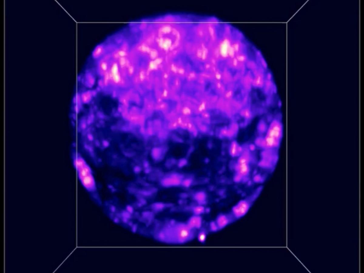
A 3D scan of an embryo created using light microscopy.
The inability to accurately predict embryo viability prior to implantation is a key contributor in the low success rate of clinical in-vitro fertilisation (IVF), but a team of experts is highlighting a safe way to study embryos using 3D optical imaging.
The team has found that the DNA damage to the embryo when using light sheet microscopy was less than when using confocal microscopy, previously considered the gold standard.
"3D imaging captures information in three dimensions: length, width, and height. To do this, microscopes will take individual images, or slices, through the embryo, which are then reconstructed," said the University of Adelaide's Associate Professor Kylie Dunning.
"At this early stage of development (this would be just prior to transfer into a patient in an IVF cycle), the embryo is made of 2 different types of cells (one that will form the foetus and the other, the placenta).
"3D imaging lets us 'see within' the embryo, capturing the health of all these cells."
Associate Professor Dunning, from the University of Adelaide's Robinson Research Institute and the Institute for Photonic and Advanced Sensing (IPAS), and Professor Kishan Dholakia, ARC Laureate Fellow and Director of the Centre of Light for Life at the University of Adelaide and researcher at the University of St Andrews, published their findings in Nature's Scientific Reports journal.
Professor Dholakia said an image using light sheet microscopy was also acquired 10 times faster than a confocal shot.
"With confocal microscopy, for every image or slice, the entire embryo is illuminated whereas with light sheet, only the slice being imaged is illuminated," he said.
"Light sheet microscopy is a standout choice for 3D imaging of live embryos, eclipsing confocal microscopy with remarkable results,"
"While it has long been believed light sheet microscopy is gentle on samples, we now have concrete evidence to support this claim for embryo imaging."
The other authors of this work were Darren JX Chow, Erik P Schartner, Stella Corsetti, Avinash Upadhya, Josephine Morizet and Frank J Gunn-Moore.
The work was supported by the UK Engineering and Physical Sciences Research Council, Australian Research Council, the National Health and Medical Research Council and the European Union's Horizon 2020 project, while Associate Professor Dunning also received funding from The Hospital Research Foundation Group and the University of Adelaide.
"We have conducted a robust comparison between light sheet and confocal microscopy, assessing the impact of volumetric imaging on photobleaching (cell autofluorescence) and phototoxicity (DNA damage)," said Professor Dholakia.
"Our next steps are to evaluate whether light sheet imaging can accurately predict embryo quality in a preclinical model."


