As fascinating as it is to work in a modern biology lab, in many cases a lot of repetitive, detailed work is necessary before the research can start. For example, cancer researchers are now capable of using hundreds or even thousands of small, lab-grown tumor samples - known as organoids - to test multiple cancer therapies, including immunotherapies, at once.
To produce organoids, researchers often need to mince a fresh tumor into small pieces by hand, using scissors to snip, snip, snip the specimen down to submillimeter size. This dissection work is tedious and yet often done by skilled - and usually overqualified - graduate students or research scientists.
Those days of drudgery may soon be past as researchers at the Stanford School of Engineering have developed two new tools that accelerate the precision cutting of tumor samples into submillimeter-scale organoids. Introducing: the microDicer (µDicer) and the microGrater (µGrater). The inventors believe these tools, not unlike familiar utensils used in kitchens everywhere to dice vegetables or grate cheese, will allow researchers to improve the consistency and quality of samples, which have direct impact on the quality of the data from downstream experiments such as measurements of drug response.
"Our collaborator and co-author Calvin Kuo, co-lead of the Cancer Biology Program at the Stanford Cancer Center, has shown that in cancer immunotherapy research, maintaining the spatial relationships between groups of tumor cells and immune cells that have already infiltrated the tumor - as opposed to working with individual cells - is really important for accuracy in testing drugs and immunotherapies. Our tools help researchers more efficiently create organoids that maintain those relationships," says Sindy Tang, an associate professor of mechanical engineering and senior author of a study appearing in the journal Microsystems & Nanoengineering describing the microDicer and the microGrater.
"These new tools will speed up the manual lab work, but their utility goes beyond that obvious advantage," Tang added. "These tools produce uniform sized organoids and the blades can be varied to whatever size the researcher requires."
Having precise control on organoid size allows researchers to ask the question whether there is a "perfect" organoid size. If the organoid is too small, it may not retain the original properties of the tumor as it existed in the body. If the organoid is too big, however, it may die due to the difficulty of oxygen and nutrients getting through to the interior tumor cells.
Precise blades
Tang and her team fashioned the blades of the microDicer in silicon using micromachining tools of classical computer chipmaking. The blade patterns are etched into silicon using a reactive plasma. The blades of the microGrater are made of stainless steel.
","additional":{"file_info":{"file_name":"SEM_1200b84.jpg","size_readable":"782.1 KB","size_bytes":800914,"width":1500,"height":1219,"modified_readable":"Aug 28, 2024 1:54 PM","modified_unix":1724878444},"varieties":[{"id":"158766:v1","type":"image_variety","type_name":"Image Variety","version":"","name":"100w","short_name":"100w","status":{"id":16,"code":"live","name":"Live"},"created":{"date":"1969-12-31T16:00:00-08:00","user_id":null},"updated":{"date":"1969-12-31T16:00:00-08:00","user_id":null},"published":{"date":null,"user_id":null},"status_changed":{"date":"1969-12-31T16:00:00-08:00","user_id":null},"urls":["https://news.stanford.edu/__data/assets/image/0033/158766/varieties/100w.jpg"],"url":"https://news.stanford.edu/__data/assets/image/0033/158766/varieties/100w.jpg","attributes":{"variety_type":"resize","filename":"100w.jpg","name":"100w","title":"microDicer and microGrater","caption":"Credit: Seth Cordts and Saisneha Koppaka","variety_size":4273,"variety_height":100,"variety_width":100,"alt":"Scanning microscopy images of the μDicer (blade spacings 200 μm, 400 μm, and 800 μm, respectively) and μGrater.","dimension_min":0,"dimension":100,"height":0,"width":0,"constrain":"centre_weighted_square"},"metadata":null,"contents":"","additional":{}},{"id":"158766:v2","type":"image_variety","type_name":"Image Variety","version":"","name":"150w","short_name":"150w","status":{"id":16,"code":"live","name":"Live"},"created":{"date":"1969-12-31T16:00:00-08:00","user_id":null},"updated":{"date":"1969-12-31T16:00:00-08:00","user_id":null},"published":{"date":null,"user_id":null},"status_changed":{"date":"1969-12-31T16:00:00-08:00","user_id":null},"urls":["https://news.stanford.edu/__data/assets/image/0033/158766/varieties/150w.jpg"],"url":"https://news.stanford.edu/__data/assets/image/0033/158766/varieties/150w.jpg","attributes":{"variety_type":"resize","filename":"150w.jpg","name":"150w","title":"microDicer and microGrater","caption":"Credit: Seth Cordts and Saisneha Koppaka","variety_size":7362,"variety_height":150,"variety_width":150,"alt":"Scanning microscopy images of the μDicer (blade spacings 200 μm, 400 μm, and 800 μm, respectively) and μGrater.","dimension_min":0,"dimension":150,"height":0,"width":0,"constrain":"centre_weighted_square"},"metadata":null,"contents":"","additional":{}},{"id":"158766:v3","type":"image_variety","type_name":"Image Variety","version":"","name":"200w","short_name":"200w","status":{"id":16,"code":"live","name":"Live"},"created":{"date":"1969-12-31T16:00:00-08:00","user_id":null},"updated":{"date":"1969-12-31T16:00:00-08:00","user_id":null},"published":{"date":null,"user_id":null},"status_changed":{"date":"1969-12-31T16:00:00-08:00","user_id":null},"urls":["https://news.stanford.edu/__data/assets/image/0033/158766/varieties/200w.jpg"],"url":"https://news.stanford.edu/__data/assets/image/0033/158766/varieties/200w.jpg","attributes":{"variety_type":"resize","filename":"200w.jpg","name":"200w","title":"microDicer and microGrater","caption":"Credit: Seth Cordts and Saisneha Koppaka","variety_size":9916,"variety_height":163,"variety_width":200,"alt":"Scanning microscopy images of the μDicer (blade spacings 200 μm, 400 μm, and 800 μm, respectively) and μGrater.","dimension_min":0,"dimension":0,"height":200,"width":200,"constrain":"width"},"metadata":null,"contents":"","additional":{}},{"id":"158766:v4","type":"image_variety","type_name":"Image Variety","version":"","name":"300w","short_name":"300w","status":{"id":16,"code":"live","name":"Live"},"created":{"date":"1969-12-31T16:00:00-08:00","user_id":null},"updated":{"date":"1969-12-31T16:00:00-08:00","user_id":null},"published":{"date":null,"user_id":null},"status_changed":{"date":"1969-12-31T16:00:00-08:00","user_id":null},"urls":["https://news.stanford.edu/__data/assets/image/0033/158766/varieties/300w.jpg"],"url":"https://news.stanford.edu/__data/assets/image/0033/158766/varieties/300w.jpg","attributes":{"variety_type":"resize","filename":"300w.jpg","name":"300w","title":"microDicer and microGrater","caption":"Credit: Seth Cordts and Saisneha Koppaka","variety_size":9916,"variety_height":163,"variety_width":200,"alt":"Scanning microscopy images of the μDicer (blade spacings 200 μm, 400 μm, and 800 μm, respectively) and μGrater.","dimension_min":0,"dimension":0,"height":300,"width":200,"constrain":"width"},"metadata":null,"contents":"","additional":{}},{"id":"158766:v5","type":"image_variety","type_name":"Image Variety","version":"","name":"345w","short_name":"345w","status":{"id":16,"code":"live","name":"Live"},"created":{"date":"1969-12-31T16:00:00-08:00","user_id":null},"updated":{"date":"1969-12-31T16:00:00-08:00","user_id":null},"published":{"date":null,"user_id":null},"status_changed":{"date":"1969-12-31T16:00:00-08:00","user_id":null},"urls":["https://news.stanford.edu/__data/assets/image/0033/158766/varieties/345w.jpg"],"url":"https://news.stanford.edu/__data/assets/image/0033/158766/varieties/345w.jpg","attributes":{"variety_type":"resize","filename":"345w.jpg","name":"345w","title":"microDicer and microGrater","caption":"Credit: Seth Cordts and Saisneha Koppaka","variety_size":24158,"variety_height":280,"variety_width":345,"alt":"Scanning microscopy images of the μDicer (blade spacings 200 μm, 400 μm, and 800 μm, respectively) and μGrater.","dimension_min":0,"dimension":0,"height":555,"width":345,"constrain":"width"},"metadata":null,"contents":"","additional":{}},{"id":"158766:v6","type":"image_variety","type_name":"Image Variety","version":"","name":"375w","short_name":"375w","status":{"id":16,"code":"live","name":"Live"},"created":{"date":"1969-12-31T16:00:00-08:00","user_id":null},"updated":{"date":"1969-12-31T16:00:00-08:00","user_id":null},"published":{"date":null,"user_id":null},"status_changed":{"date":"1969-12-31T16:00:00-08:00","user_id":null},"urls":["https://news.stanford.edu/__data/assets/image/0033/158766/varieties/375w.jpg"],"url":"https://news.stanford.edu/__data/assets/image/0033/158766/varieties/375w.jpg","attributes":{"variety_type":"resize","filename":"375w.jpg","name":"375w","title":"microDicer and microGrater","caption":"Credit: Seth Cordts and Saisneha Koppaka","variety_size":28083,"variety_height":305,"variety_width":375,"alt":"Scanning microscopy images of the μDicer (blade spacings 200 μm, 400 μm, and 800 μm, respectively) and μGrater.","dimension_min":0,"dimension":0,"height":200,"width":375,"constrain":"width"},"metadata":null,"contents":"","additional":{}},{"id":"158766:v7","type":"image_variety","type_name":"Image Variety","version":"","name":"400w","short_name":"400w","status":{"id":16,"code":"live","name":"Live"},"created":{"date":"1969-12-31T16:00:00-08:00","user_id":null},"updated":{"date":"1969-12-31T16:00:00-08:00","user_id":null},"published":{"date":null,"user_id":null},"status_changed":{"date":"1969-12-31T16:00:00-08:00","user_id":null},"urls":["https://news.stanford.edu/__data/assets/image/0033/158766/varieties/400w.jpg"],"url":"https://news.stanford.edu/__data/assets/image/0033/158766/varieties/400w.jpg","attributes":{"variety_type":"resize","filename":"400w.jpg","name":"400w","title":"microDicer and microGrater","caption":"Credit: Seth Cordts and Saisneha Koppaka","variety_size":30872,"variety_height":325,"variety_width":400,"alt":"Scanning microscopy images of the μDicer (blade spacings 200 μm, 400 μm, and 800 μm, respectively) and μGrater.","dimension_min":0,"dimension":0,"height":100,"width":400,"constrain":"width"},"metadata":null,"contents":"","additional":{}},{"id":"158766:v8","type":"image_variety","type_name":"Image Variety","version":"","name":"555w","short_name":"555w","status":{"id":16,"code":"live","name":"Live"},"created":{"date":"1969-12-31T16:00:00-08:00","user_id":null},"updated":{"date":"1969-12-31T16:00:00-08:00","user_id":null},"published":{"date":null,"user_id":null},"status_changed":{"date":"1969-12-31T16:00:00-08:00","user_id":null},"urls":["https://news.stanford.edu/__data/assets/image/0033/158766/varieties/555w.jpg"],"url":"https://news.stanford.edu/__data/assets/image/0033/158766/varieties/555w.jpg","attributes":{"variety_type":"resize","filename":"555w.jpg","name":"555w","title":"microDicer and microGrater","caption":"Credit: Seth Cordts and Saisneha Koppaka","variety_size":54008,"variety_height":451,"variety_width":555,"alt":"Scanning microscopy images of the μDicer (blade spacings 200 μm, 400 μm, and 800 μm, respectively) and μGrater.","dimension_min":0,"dimension":0,"height":0,"width":555,"constrain":"width"},"metadata":null,"contents":"","additional":{}},{"id":"158766:v9","type":"image_variety","type_name":"Image Variety","version":"","name":"600w","short_name":"600w","status":{"id":16,"code":"live","name":"Live"},"created":{"date":"1969-12-31T16:00:00-08:00","user_id":null},"updated":{"date":"1969-12-31T16:00:00-08:00","user_id":null},"published":{"date":null,"user_id":null},"status_changed":{"date":"1969-12-31T16:00:00-08:00","user_id":null},"urls":["https://news.stanford.edu/__data/assets/image/0033/158766/varieties/600w.jpg"],"url":"https://news.stanford.edu/__data/assets/image/0033/158766/varieties/600w.jpg","attributes":{"variety_type":"resize","filename":"600w.jpg","name":"600w","title":"microDicer and microGrater","caption":"Credit: Seth Cordts and Saisneha Koppaka","variety_size":61706,"variety_height":488,"variety_width":600,"alt":"Scanning microscopy images of the μDicer (blade spacings 200 μm, 400 μm, and 800 μm, respectively) and μGrater.","dimension_min":0,"dimension":0,"height":0,"width":600,"constrain":"width"},"metadata":null,"contents":"","additional":{}},{"id":"158766:v10","type":"image_variety","type_name":"Image Variety","version":"","name":"690w","short_name":"690w","status":{"id":16,"code":"live","name":"Live"},"created":{"date":"1969-12-31T16:00:00-08:00","user_id":null},"updated":{"date":"1969-12-31T16:00:00-08:00","user_id":null},"published":{"date":null,"user_id":null},"status_changed":{"date":"1969-12-31T16:00:00-08:00","user_id":null},"urls":["https://news.stanford.edu/__data/assets/image/0033/158766/varieties/690w.jpg"],"url":"https://news.stanford.edu/__data/assets/image/0033/158766/varieties/690w.jpg","attributes":{"variety_type":"resize","filename":"690w.jpg","name":"690w","title":"microDicer and microGrater","caption":"Credit: Seth Cordts and Saisneha Koppaka","variety_size":78472,"variety_height":561,"variety_width":690,"alt":"Scanning microscopy images of the μDicer (blade spacings 200 μm, 400 μm, and 800 μm, respectively) and μGrater.","dimension_min":0,"dimension":0,"height":0,"width":690,"constrain":"width"},"metadata":null,"contents":"","additional":{}},{"id":"158766:v11","type":"image_variety","type_name":"Image Variety","version":"","name":"705w","short_name":"705w","status":{"id":16,"code":"live","name":"Live"},"created":{"date":"1969-12-31T16:00:00-08:00","user_id":null},"updated":{"date":"1969-12-31T16:00:00-08:00","user_id":null},"published":{"date":null,"user_id":null},"status_changed":{"date":"1969-12-31T16:00:00-08:00","user_id":null},"urls":["https://news.stanford.edu/__data/assets/image/0033/158766/varieties/705w.jpg"],"url":"https://news.stanford.edu/__data/assets/image/0033/158766/varieties/705w.jpg","attributes":{"variety_type":"resize","filename":"705w.jpg","name":"705w","title":"microDicer and microGrater","caption":"Credit: Seth Cordts and Saisneha Koppaka","variety_size":81970,"variety_height":573,"variety_width":705,"alt":"Scanning microscopy images of the μDicer (blade spacings 200 μm, 400 μm, and 800 μm, respectively) and μGrater.","dimension_min":0,"dimension":0,"height":0,"width":705,"constrain":"width"},"metadata":null,"contents":"","additional":{}},{"id":"158766:v12","type":"image_variety","type_name":"Image Variety","version":"","name":"750w","short_name":"750w","status":{"id":16,"code":"live","name":"Live"},"created":{"date":"1969-12-31T16:00:00-08:00","user_id":null},"updated":{"date":"1969-12-31T16:00:00-08:00","user_id":null},"published":{"date":null,"user_id":null},"status_changed":{"date":"1969-12-31T16:00:00-08:00","user_id":null},"urls":["https://news.stanford.edu/__data/assets/image/0033/158766/varieties/750w.jpg"],"url":"https://news.stanford.edu/__data/assets/image/0033/158766/varieties/750w.jpg","attributes":{"variety_type":"resize","filename":"750w.jpg","name":"750w","title":"microDicer and microGrater","caption":"Credit: Seth Cordts and Saisneha Koppaka","variety_size":91742,"variety_height":610,"variety_width":750,"alt":"Scanning microscopy images of the μDicer (blade spacings 200 μm, 400 μm, and 800 μm, respectively) and μGrater.","dimension_min":0,"dimension":0,"height":0,"width":750,"constrain":"width"},"metadata":null,"contents":"","additional":{}},{"id":"158766:v13","type":"image_variety","type_name":"Image Variety","version":"","name":"795w","short_name":"795w","status":{"id":16,"code":"live","name":"Live"},"created":{"date":"1969-12-31T16:00:00-08:00","user_id":null},"updated":{"date":"1969-12-31T16:00:00-08:00","user_id":null},"published":{"date":null,"user_id":null},"status_changed":{"date":"1969-12-31T16:00:00-08:00","user_id":null},"urls":["https://news.stanford.edu/__data/assets/image/0033/158766/varieties/795w.jpg"],"url":"https://news.stanford.edu/__data/assets/image/0033/158766/varieties/795w.jpg","attributes":{"variety_type":"resize","filename":"795w.jpg","name":"795w","title":"microDicer and microGrater","caption":"Credit: Seth Cordts and Saisneha Koppaka","variety_size":98538,"variety_height":646,"variety_width":795,"alt":"Scanning microscopy images of the μDicer (blade spacings 200 μm, 400 μm, and 800 μm, respectively) and μGrater.","dimension_min":0,"dimension":0,"height":0,"width":795,"constrain":"width"},"metadata":null,"contents":"","additional":{}},{"id":"158766:v14","type":"image_variety","type_name":"Image Variety","version":"","name":"960w","short_name":"960w","status":{"id":16,"code":"live","name":"Live"},"created":{"date":"1969-12-31T16:00:00-08:00","user_id":null},"updated":{"date":"1969-12-31T16:00:00-08:00","user_id":null},"published":{"date":null,"user_id":null},"status_changed":{"date":"1969-12-31T16:00:00-08:00","user_id":null},"urls":["https://news.stanford.edu/__data/assets/image/0033/158766/varieties/960w.jpg"],"url":"https://news.stanford.edu/__data/assets/image/0033/158766/varieties/960w.jpg","attributes":{"variety_type":"resize","filename":"960w.jpg","name":"960w","title":"microDicer and microGrater","caption":"Credit: Seth Cordts and Saisneha Koppaka","variety_size":135716,"variety_height":780,"variety_width":960,"alt":"Scanning microscopy images of the μDicer (blade spacings 200 μm, 400 μm, and 800 μm, respectively) and μGrater.","dimension_min":0,"dimension":0,"height":0,"width":960,"constrain":"width"},"metadata":null,"contents":"","additional":{}},{"id":"158766:v15","type":"image_variety","type_name":"Image Variety","version":"","name":"1024w","short_name":"1024w","status":{"id":16,"code":"live","name":"Live"},"created":{"date":"1969-12-31T16:00:00-08:00","user_id":null},"updated":{"date":"1969-12-31T16:00:00-08:00","user_id":null},"published":{"date":null,"user_id":null},"status_changed":{"date":"1969-12-31T16:00:00-08:00","user_id":null},"urls":["https://news.stanford.edu/__data/assets/image/0033/158766/varieties/1024w.jpg"],"url":"https://news.stanford.edu/__data/assets/image/0033/158766/varieties/1024w.jpg","attributes":{"variety_type":"resize","filename":"1024w.jpg","name":"1024w","title":"microDicer and microGrater","caption":"Credit: Seth Cordts and Saisneha Koppaka","variety_size":149115,"variety_height":832,"variety_width":1024,"alt":"Scanning microscopy images of the μDicer (blade spacings 200 μm, 400 μm, and 800 μm, respectively) and μGrater.","dimension_min":0,"dimension":0,"height":0,"width":1024,"constrain":"width"},"metadata":null,"contents":"","additional":{}},{"id":"158766:v16","type":"image_variety","type_name":"Image Variety","version":"","name":"1110w","short_name":"1110w","status":{"id":16,"code":"live","name":"Live"},"created":{"date":"1969-12-31T16:00:00-08:00","user_id":null},"updated":{"date":"1969-12-31T16:00:00-08:00","user_id":null},"published":{"date":null,"user_id":null},"status_changed":{"date":"1969-12-31T16:00:00-08:00","user_id":null},"urls":["https://news.stanford.edu/__data/assets/image/0033/158766/varieties/1110w.jpg"],"url":"https://news.stanford.edu/__data/assets/image/0033/158766/varieties/1110w.jpg","attributes":{"variety_type":"resize","filename":"1110w.jpg","name":"1110w","title":"microDicer and microGrater","caption":"Credit: Seth Cordts and Saisneha Koppaka","variety_size":172185,"variety_height":902,"variety_width":1110,"alt":"Scanning microscopy images of the μDicer (blade spacings 200 μm, 400 μm, and 800 μm, respectively) and μGrater.","dimension_min":0,"dimension":0,"height":0,"width":1110,"constrain":"width"},"metadata":null,"contents":"","additional":{}},{"id":"158766:v17","type":"image_variety","type_name":"Image Variety","version":"","name":"1410w","short_name":"1410w","status":{"id":16,"code":"live","name":"Live"},"created":{"date":"1969-12-31T16:00:00-08:00","user_id":null},"updated":{"date":"1969-12-31T16:00:00-08:00","user_id":null},"published":{"date":null,"user_id":null},"status_changed":{"date":"1969-12-31T16:00:00-08:00","user_id":null},"urls":["https://news.stanford.edu/__data/assets/image/0033/158766/varieties/1410w.jpg"],"url":"https://news.stanford.edu/__data/assets/image/0033/158766/varieties/1410w.jpg","attributes":{"variety_type":"resize","filename":"1410w.jpg","name":"1410w","title":"microDicer and microGrater","caption":"Credit: Seth Cordts and Saisneha Koppaka","variety_size":250521,"variety_height":1146,"variety_width":1410,"alt":"Scanning microscopy images of the μDicer (blade spacings 200 μm, 400 μm, and 800 μm, respectively) and μGrater.","dimension_min":0,"dimension":0,"height":0,"width":1410,"constrain":"width"},"metadata":null,"contents":"","additional":{}},{"id":"158766:v18","type":"image_variety","type_name":"Image Variety","version":"","name":"1500w","short_name":"1500w","status":{"id":16,"code":"live","name":"Live"},"created":{"date":"1969-12-31T16:00:00-08:00","user_id":null},"updated":{"date":"1969-12-31T16:00:00-08:00","user_id":null},"published":{"date":null,"user_id":null},"status_changed":{"date":"1969-12-31T16:00:00-08:00","user_id":null},"urls":["https://news.stanford.edu/__data/assets/image/0033/158766/varieties/1500w.jpg"],"url":"https://news.stanford.edu/__data/assets/image/0033/158766/varieties/1500w.jpg","attributes":{"variety_type":"resize","filename":"1500w.jpg","name":"1500w","title":"microDicer and microGrater","caption":"Credit: Seth Cordts and Saisneha Koppaka","variety_size":299928,"variety_height":1219,"variety_width":1500,"alt":"Scanning microscopy images of the μDicer (blade spacings 200 μm, 400 μm, and 800 μm, respectively) and μGrater.","dimension_min":0,"dimension":0,"height":0,"width":1500,"constrain":"width"},"metadata":null,"contents":"","additional":{}}],"exif":{"image":{"ImageHeight":1219,"ImageWidth":1500,"ModifyDate":"2024:08:28 13:25:48","Orientation":0,"ResolutionUnit":0,"Software":"Adobe Photoshop 24.4 (Macintosh)","XResolution":1200,"YResolution":1200},"thumbnail":{"Compression":0,"ThumbnailLength":3583,"ThumbnailOffset":318},"exif":{"ColorSpace":0,"CreateDate":"2024:08:28 13:18:01-07:00","ExifImageHeight":1219,"ExifImageWidth":1500}}}}}">
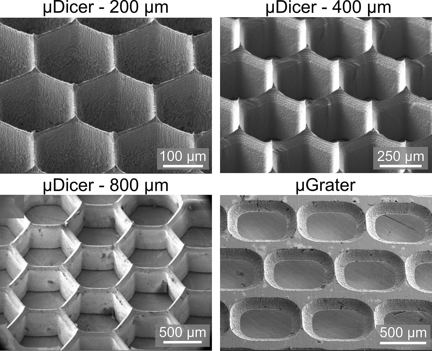
Scanning microscopy images of the microDicer (blade spacings 200 μm, 400 μm, and 800 μm, respectively) and microGrater. | Seth Cordts and Saisneha Koppaka


