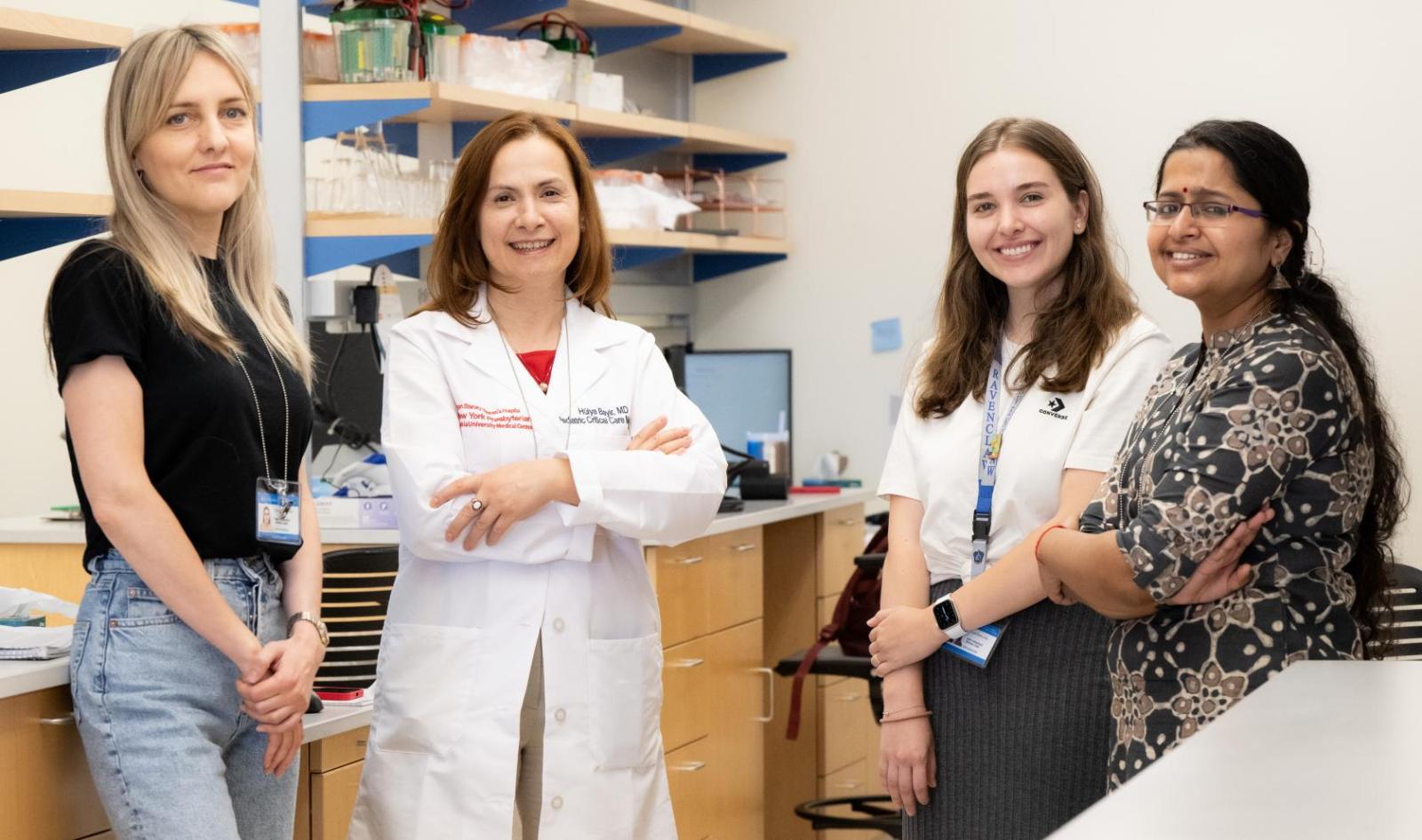
Each year close to half a million children who suffer falls, assaults, motor vehicle or bicycle accidents, or sports injuries are admitted to emergency rooms in the US with a traumatic brain injury (TBI)-and among all types of injuries in young people, TBIs are most likely to cause permanent disability or death. Columbia's recently appointed chief of pediatric critical care and hospital medicine, Hülya Bayır, MD, has dedicated her career to understanding the brain's special vulnerability to injury, and what might be done to protect brain tissue in the hours and days after an accident. Through the newly established Columbia Children's Redox Health Center, she and colleagues are identifying and characterizing a set of key cellular changes that occur following a brain injury, called redox reactions-shorthand for reduction and oxidation.
"To quote one of my mentors, in pediatrics we treat 20 percent of the population, but they are 100 percent of the future."
Redox reactions are fundamental to all life: during plant photosynthesis, for example, carbon dioxide is reduced into sugars, and water is oxidized into molecular oxygen. These reactions also take place continuously throughout the human body and are essential for energy production, fighting infections, and for learning and memory. But in some contexts they can also have destructive effects, Dr. Bayır explains. "Many people know the products of these reactions as free radicals, reactive oxygen species, and oxidative stress, which have been implicated in aging and in almost every disease. Our goal is to understand exactly what the products of these redox reactions are, and how their normal processes go bad."
A healthy brain is a marvel of energy production, using about 20 percent of both the body's oxygen and glucose supplies in highly regulated metabolic reactions that generate energy to maintain central nervous system function. In an injured brain these reactions can become uncontrolled, leading to a cascade of damaging processes including dysregulation of the brain's blood flow, damage to the blood brain barrier (which protects the brain from toxins), and an activation of inflammatory responses and programmed cell death pathways.
Brain tissue is rich with fatty compounds called lipids, building blocks in razor-thin structures called biomembranes, which separate cells, and organelles inside cells, from each other. These membranes also help regulate and coordinate myriad metabolic processes that occur in the brain.

Dr. Bayır and her lab team work to understand the brain's special vulnerability to injury, and what might be done to protect brain tissue in the hours and days after an accident.
In response to a brain injury, brain lipids may undergo a redox process called "lipid peroxidation." Lipids are transformed into unstable lipid hydroperoxides, which then readily break down into multiple secondary products that can trigger the subsequent cellular injuries that occur after TBI. Lipid peroxidation is also a factor in neurological disorders including Alzheimer's and Parkinson's diseases, stroke, and Down syndrome.
Collaborating with chemists, biophysicists, analytical chemists, and using highly advanced and rapidly evolving tools such as imaging mass spectrometry, Dr. Bayır is developing methods to identify lipid peroxidation products, and to compile them into a sort of "dictionary" that shows the role these products play in cellular communications, and whether they're damaging or beneficial.
The lipid language of cells is quite complex, Dr. Bayır notes. "We've been able to identify what some of these molecules are communicating; we now know how they signal 'survive' and 'die,' or 'this cell must be removed,'" she says.
Brain cells that are injured beyond repair are destined to die through one or more processes called regulated cell death, which are critical to containing the injury and protecting the entire cell community. Since the first programmed cell death pathway, apoptosis, was described in 1965, researchers have identified the oxidized lipid signals in more than 30 different programmed cell death pathways. (One of those pathways, called ferroptosis, was identified here at Columbia in 2012 by Brent Stockwell, PhD and his team.)
Dr. Bayır's research has major implications for diseases and disorders in addition to TBI. The mass spectrometry method her team has developed enables researchers to image proteins, lipids, metabolites in the same tissue, and understand their spatial organization and relationship to each other. This is key to understanding how tumor cells alter the microenvironments around them, and how they generate signals that suppress the immune function of neighboring cells.
Her research also could have implications for patients with stroke, cardiac arrest, sepsis (overwhelming infection of the body), acute kidney injury, or radiation damage that can occur when normal tissue is exposed during radiotherapy treatment for cancer, as well as cellular damage caused by a nuclear accident or exposure to a dirty bomb.
Dr. Bayır's overarching goal is to design novel drug therapies or identify existing, potentially effective medications that could not only save essential brain cells from destruction, but that might also apply to other diseases. This goal is especially motivating when caring for children who have the misfortune to land in the intensive care unit, she says. "To quote one of my mentors, in pediatrics we treat 20 percent of the population, but they are 100 percent of the future."


