ETH Zurich researchers have investigated how microbubbles tiny gas bubbles can deliver drugs into cells in a targeted manner using ultrasound. For the first time, they have visualised how tiny cyclic microjets liquid jets generated by microbubbles penetrate the cell membrane enabling the drug uptake.
In brief
- ETH Zurich researchers show how under ultrasound, microbubbles gas-filled bubbles generate liquid jets that penetrate cell membrane like tiny pinpricks.
- The dynamics of jets is governed by a hydrodynamic instability which can be controlled precisely using different combinations of ultrasound frequency, pressure, and microbubble size.
- Explanation of mechanism behind drug delivery via microbubbles lays important foundations for transporting drugs into the brain, for example.
The targeted treatment of brain diseases such as Alzheimer's, Parkinson's or brain tumours is challenging because the brain is a particularly sensitive organ that is well protected. That's why researchers are working on ways of delivering drugs to the brain precisely, via the bloodstream. The aim is to overcome the blood-brain barrier which normally only allows certain nutrients and oxygen to pass through.
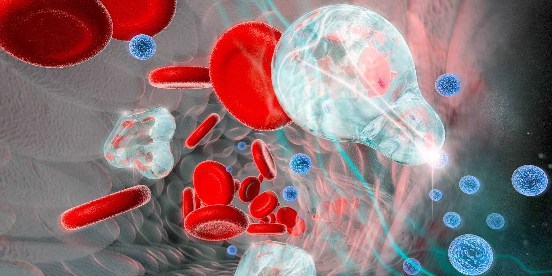
Microbubbles that react to ultrasound are a particularly promising method for this sort of therapy. These microbubbles are smaller than a red blood cell, filled with gas and have consist of a special coating of fat molecules to stabilise them. They are injected into the bloodstream together with the drug and then activated at the target site using ultrasound. The action movement of the microbubbles creates tiny pores in the cell membrane of the blood vessel wall which the drug can then pass through.
How exactly microbubbles create these pores was previously unclear. Now, a group of ETH researchers, led by Outi Supponen, a professor at the Institute of Fluid Dynamics, have been able to demonstrate for the first time how this mechanism works. "We were able to show that under ultrasound, the surface of the microbubbles loses theirits shape, resulting in tiny jets of liquid, so-called microjets, which penetrate the cell membrane," explains Marco Cattaneo, Supponen's doctoral student and lead author of the study which was recently published in Nature Physics.
The invisible force: liquid microjets at 200 kph
Until now, nobody knew how the pores in the cell membrane were formed because the microbubbles measure just a few micrometres across and vibrate up to several million times per second under ultrasound. This is a process that is incredibly difficult to observe and which requires a special set-up in the lab. "So far, most studies have looked at the process from above through a conventional microscope. But when you do that, you can't see what's happening between the microbubble and the cell," says Cattaneo. The researchers therefore built a microscope with a magnification of 200x, which allows them to observe the process from the side, and connected it to a high-speed camera that can take up to ten million images per second.
For their experiment, they mimicked the blood vessel wall using an in-vitro model, growing endothelial cells on a plastic membrane. They placed this membrane on a box with transparent walls filled with a saline solution and a model drug, with the cells facing down like a lid. The gas-filled microbubble rose to the top automatically and made contact with the cells. The microbubbles were then set in vibration by a microsecond-long pulse of ultrasound.

"At a sufficiently high ultrasound pressure, microbubbles stop oscillating in a spherical shape and start reshaping themselves into regular, non-spherical patterns," says Supponen. The "lobes" of these patterns oscillate cyclically, pushing inwards and outwards. The researchers discovered that above a certain ultrasound pressure, the inward-folded lobes can become so deeply sunken that they generate powerful jets, crossing the entire bubble and making contact with the cell.
These microjets move at an incredible speed of 200 kph and are able to perforate the cell membrane like a targeted pinprick without destroying the cell. This jet mechanism does not destroy the bubble, meaning that a new microjet can form with each ultrasound cycle.
Physics in the service of medicine
"An intriguing aspect is that this ejection mechanism is triggered at low ultrasound pressures, around 100 kPa," says Supponen. This means that the ultrasound pressure acting on the microbubbles, and therefore on the patient, is comparable to the atmospheric air pressure that is around us all the time.
The researchers from Supponen's group not only made visual observations, but also provided explanations using a range of different theoretical models. They were able to show that the microjets have by far the greatest potential for damage over the many other mechanisms that have been proposed in the past, strongly supporting the researchers' observation that the cell membrane is pierced only when a microjet is generated. Cattaneo says, "With our lab setup, we now have a better way of observing the microbubbles and can describe the cell-microbubble interaction more precisely." This system can also be used to investigate how new microbubble formulations developed by other researchers react to ultrasound, for example.
Supponen adds, "Our work clarifies the physical foundations for targeted administration of drugs through microbubbles and helps us define criteria for their safe and effective use." This means that the right combination of frequency, pressure and microbubble size can help to maximise the outcome of the therapy, while ensuring greater safety and lower risk to patients. "Additionally, we were able to show that just a few pulses of ultrasound are enough to perforate a cell membrane. This is also good news for patients," says Supponen. Conversely, the coating of the microbubbles can also be optimised for the required ultrasound frequency, making it easier for the jets to form.
Reference
Cattaneo M, Guerriero G, Gazendra S, Krattiger L, Paganella L, Narciso M, Supponen O, Cyclic jetting enables microbubble-mediated drug delivery, Nature Physics (2025), DOI: external page https://doi.org/10.1038/s41567-025-02785-0


