Another EMBL-engineered advance to Brillouin microscopy significantly widens the aperture and provides for quick, more efficient 3-D imaging of light-sensitive samples
Summary
- Previously, EMBL scientists developed a new microscope based on Brillouin scattering - a phenomenon where light interacts with naturally occurring thermal vibrations within materials, from which their mechanical properties can be deduced. This method has since been used for non-invasive and live-imaging applications in biology, and that advance was selected as one of The Guardian's 10 biggest science stories of 2022 .
- In a new paper, the same scientists have now succeeded in substantially further advancing Brillouin microscopy, making it approximately a thousand times faster and more efficient.
- Simultaneously, the new microscopy method expands the swath of material it can view - from a 100-pixel line to a ~10,000-pixel full plane, capturing 3D images quickly enough for live-organism observation.
A new paper updates an EMBL technology advance even further. More details about the original technology can be found in our initial reporting here .
EMBL tech developers have made an important leap forward with a novel methodology that adds an important microscopy capability to life scientists' toolbox. The advance represents a 1,000-fold improvement in speed and throughput in Brillouin microscopy and provides a way to view light-sensitive organisms more efficiently.
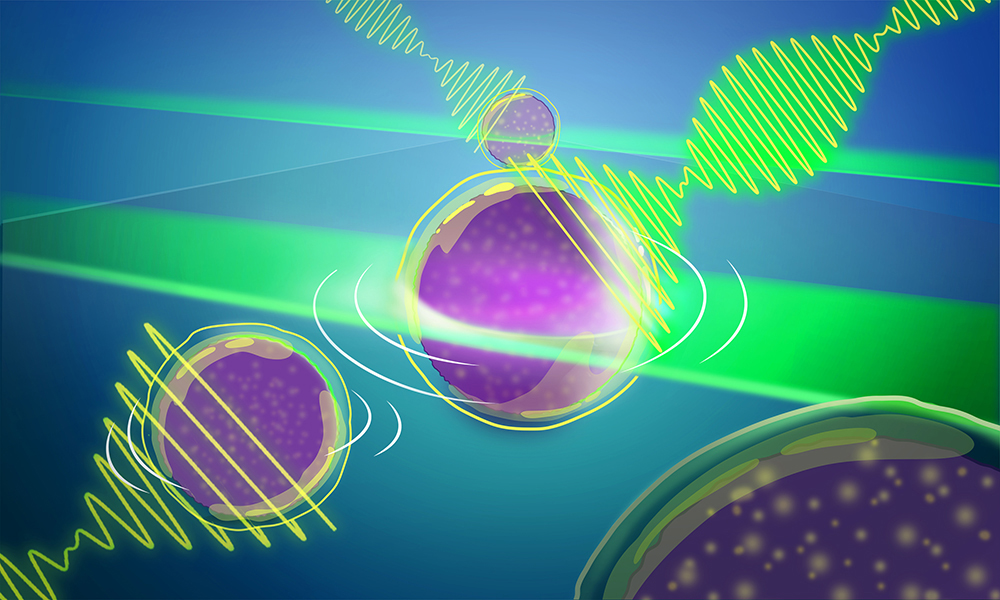
"We were on a quest to speed up image acquisition," said Carlo Bevilacqua, lead author on a paper published about this technological development in Nature Photonics and an optical engineer in EMBL's Prevedel Team. "Over the years, we have progressed from being able to see just a pixel at a time to a line of 100 pixels, to now a full plane that offers a view of approximately 10,000 pixels."
The technology is based on a phenomenon first predicted in 1922 by French physicist Léon Brillouin. He showed that when light is shone on a material, it interacts with naturally occurring thermal vibrations within, exchanging energy and thereby slightly shifting the frequency (or colour) of the light. Measuring the spectrum (colours) of the scattered light reveals information about a material's physical characteristics.
Using Brillouin scattering for microscopy purposes came much later - in the early 2000s - when other technological advancements enabled scientists to measure tiny frequency shifts with high precision and sufficient throughput. This allowed them to compute mechanical properties of living biological samples. However, at that point, scientists were only able to view one pixel at a time. The process was therefore quite time-consuming, and it severely limited how the microscopy method could be used in biology. In 2022, Bevilacqua and others in the Prevedel group were able to first expand the field of view to a line, and now with this latest development, to a full 2D field of view, which also helps speed up 3D imaging.
"Just as the development of light-sheet microscopy here at EMBL marked a revolution in light microscopy because it allowed for faster, high-resolution, and minimally phototoxic imaging of biological samples, so too does this advance in the area of mechanical or Brillouin imaging," said Robert Prevedel, Group Leader and senior author on the paper. "We hope this new technology - with minimal light intensity - opens one more 'window' for life scientists' exploration."


