Using a new method to study how carbohydrates modify proteins, scientists have discovered that gut bacteria can alter molecular signatures in the brain
Summary
- Glycosylation is the process by which cells add sugar groups (also called carbohydrates) to proteins to modify their functions.
- EMBL researchers developed a new method to systematically and quantitatively study glycosylation and used it to show that gut bacteria can influence glycosylation patterns in the brains of mice.
- Using this method, the researchers could identify over 150,000 glycosylated forms of proteins ('proteoforms') in the brain, a more than 25-fold increase over previous studies.
- The study sheds new light on connections between the microbiome and the nervous system and provides a new method to study glycosylation's role in fundamental biological processes.
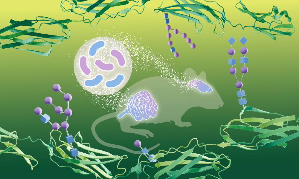
Our guts are home to trillions of bacteria, and research over the last few decades has established how essential they are to our physiology - in health and disease . A new study from EMBL Heidelberg researchers shows that gut bacteria can bring about profound molecular changes in one of our most critical organs - the brain.
The new study, published in the journal Nature Structural and Molecular Biology, is the first to show that bacteria living in the gut can influence how proteins in the brain are modified by carbohydrates - a process called glycosylation. The study was made possible by a new method the scientists developed - DQGlyco - which allows them to study glycosylation at a much higher scale and resolution than previous studies.
A new way to measure glycosylation
Proteins are the workhorses of our cells and their main building blocks. Sugars, or carbohydrates, on the other hand, are among the body's main sources of energy. However, the cell also uses sugars to chemically modify proteins, altering their functions. This is called glycosylation.
"Glycosylation can affect how cells attach to each other (adhesion), how they move (motility), and even how they talk to one another (communication)," explained Clément Potel, first author of the study and Savitski Team Research Scientist. "It is involved in the pathogenesis of several diseases, including cancer and neuronal disorders."
However, glycosylation has traditionally been notoriously difficult to study. Only a small portion of proteins in the cell are glycosylated and concentrating enough of them in a sample for studying (a process called 'enriching') tends to be laborious, expensive, and time-consuming.
"So far, it's not been possible to do such studies on a systematic scale, in a quantitative fashion, and with high reproducibility," said Mikhail Savitski, Team Leader, Senior Scientist, and Head of the Proteomics Core Facility at EMBL Heidelberg. "These are the challenges we managed to overcome with the new method."
DQGlyco uses easily available and low-cost laboratory materials, such as functionalised silica beads, to selectively enrich glycosylated proteins from biological samples, which can then be precisely identified and measured. Applying the method to brain tissue samples from mice, the researchers could identify over 150,000 glycosylated forms of proteins ('proteoforms'), an increase of over 25-fold compared to previous studies.
The quantitative nature of the new method means that researchers can compare and measure differences between samples from different tissues, cell lines, species, etc. This also lets them study the pattern of 'microheterogeneity' - the phenomenon where the same part of a protein can be modified by many (sometimes hundreds of) different sugar groups.
One of the most common examples of microheterogeneity is human blood groups, where the presence of different sugar groups on proteins in red blood cells determines blood type (A, B, O, and AB). This plays a major role in deciding the success of blood transfusions from one individual to the other.
The new method allowed the team to identify such microheterogeneity across hundreds of protein sites. "I think the widespread prevalence of microheterogeneity is something people had always assumed but had never been clearly demonstrated, since you need to have enough coverage of glycosylated proteins to be able to make the statement," said Mira Burtscher, another first author of the study and a Savitski Team PhD student.
From the gut to the brain
Given the method's precision and power, the researchers decided to use it to address an outstanding biological question. In collaboration with Michael Zimmerman's group at EMBL, they next tested whether the gut microbiome had any effect on the glycosylation signatures they had observed in the brain. Both Zimmermann and Savitski are part of the Microbial Ecosystems Transversal Theme at EMBL, which was introduced by the 2022-26 EMBL programme 'Molecules to Ecosystems'.
"It is known that gut microbiomes can affect neural functions, but the molecular details are largely unknown," said Potel. "Glycosylation is implicated in many processes, such as neurotransmission and axon guidance, so we wanted to test if this was a mechanism by which gut bacteria influenced molecular pathways in the brain."
Interestingly, the team found that when compared to 'germ-free mice', i.e. mice grown in special environments such that they completely lack a gut microbiome, mice with different complements of gut bacteria had different glycosylation patterns in the brain. The changed patterns were particularly apparent in proteins known to be important in neural functions, such as cognitive processing and axon growth.
The study's datasets are openly available via a new dedicated app for other researchers. In addition, the team is also curious whether the data can be used to inform predictions about glycosylation sites, especially in different species. For this, they have been using machine learning approaches such as AlphaFold - the AI-based tool for predicting protein structures recognised with the 2024 Nobel Prize in Chemistry.
"By training the models on mouse data, we can start predicting what could be the variability of glycosylation sites in humans, for example," said Martin Garrido, a postdoc in the Savitski and Saez-Rodriguez groups at EMBL and another first author of the study. "It could be very useful for people studying other organisms to help them identify glycosylation sites in their proteins of interest."
The researchers are also working towards applying the new method to answer more fundamental biological questions and to understand the functional role glycosylation plays in cells.


