What do fish, chameleons, crabs and Walter White, the chemistry teacher from Breaking Bad, all have in common? The answer is that they all know how to make crystals. But, unlike the incorrigible White, who manufactured mind-altering methamphetamine crystals for criminal ends, the others make natural crystals for far more wholesome purposes: from vision and camouflage to regulating heat and communicating.
Although many of the crystals that living creatures produce inside their cells are comprised of just two molecules - guanine and hypoxanthine - the resultant diversity is mind-boggling in terms of shapes, uses and optical properties. Researchers have been fascinated by animals' ability to create such huge variety from two simple building blocks, but until now there was no satisfactory explanation for this wealth of crystals in the animal world. In a study published recently in Nature Chemical Biology, Weizmann Institute of Science researchers solved the mystery and provided a comprehensive description of the "biological recipes" that cells use to cook up such a varied and useful menu of crystals.
The hero of the story is the zebrafish, a small, visually stunning freshwater fish whose body is covered with colorful crystals. Close-up photos reveal that its tissues contain different crystals: While the gills' protective cover, the operculum, is silvery, the eyes reflect a bluish light, and the skin cells are a sparkling yellow or blue.
""Zebrafish crystals provide a perfect opportunity to study how crystal structure and properties are biochemically and genetically controlled"
"While trying to understand why the crystals are different, we isolated them from the tissues and discovered that they differed greatly from one another in terms of shape, composition and arrangement in the cell," explains Dr. Dvir Gur of Weizmann's Molecular Genetics Department, who headed the research team.
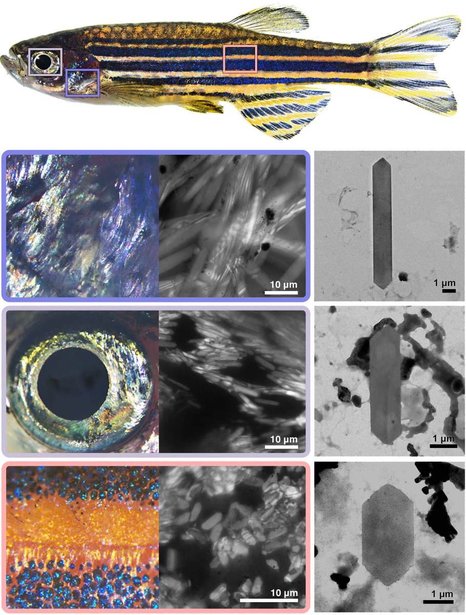
Using an electron microscope, the team observed that the crystals forming in the gills' cover were long and narrow, while those in the eyes were shorter, and the crystals in the skin were the shortest. "We realized that the zebrafish's crystals provide us with a perfect research opportunity to understand how the structure and properties of crystals are biochemically and genetically controlled, without having to compare between crystals formed in different animals with different genetic makeups," Gur adds.
When the researchers took a closer look at the zebrafish's different crystals, they noticed that their shape was affected by the ratio between the molecules of guanine and hypoxanthine. This effect resembles what happens in the work of a baker, who can choose an appropriate balance of ingredients to create different delicacies: More cream than chocolate, for example, will produce an airy mousse, while using a balanced or lower ratio will produce a ganache that is perfect for filling or glazing. Similarly, different ratios of guanine and hypoxanthine in the crystal will alter its structure and its optical properties and, accordingly, the way in which the organism uses it. After measuring the ratio of these molecular building blocks in different fish crystals, the researchers managed to artificially recreate three different zebrafish crystals in the laboratory, each containing different ratios of guanine and hypoxanthine.
The proteins that put the light back in the gills
But guanine and hypoxanthine are not just the building blocks of crystals. They are also vital molecules that all living creatures use and that are essential to both the building of DNA and the proper functioning of cells. To better understand the mechanism that regulates their ratio in crystals, and why the production of these crystals does not disrupt other cellular processes, Rachel Deis, the PhD student who led the research, isolated the crystal-producing cells, called iridophores. She then, in cooperation with an interdisciplinary research team, deciphered the biological mechanisms that enable them to create bespoke crystals.
"This is the first time that we succeeded in isolating the iridophores, identifying the proteins they contain and comparing them to cells that do not produce crystals," Deis says. "When we looked at the list of proteins in the iridophores, we were surprised to discover two apparently contradictory trends. On the one hand, the iridophores contained a particularly large quantity of enzymes responsible for forming the crystals' building blocks; on the other hand, they contained lower quantities than expected of other enzymes with similar functions that belong to the same family of proteins."
These findings led the researchers to suspect that each group of iridophores has a unique balance of enzymes, and that the unique balance in the cell is what determines the ratio between each crystal's building blocks and, as a result, the structure and function of the crystal in different tissues. "Humans have only one enzyme that is responsible for the final stage of preparing guanine; in fish, we identified five different enzymes," Gur explains. This multitude of enzymes in fish allows them to produce different ratios of guanine and hypoxanthine, the building blocks used to form the crystals, without disrupting other cellular functions.
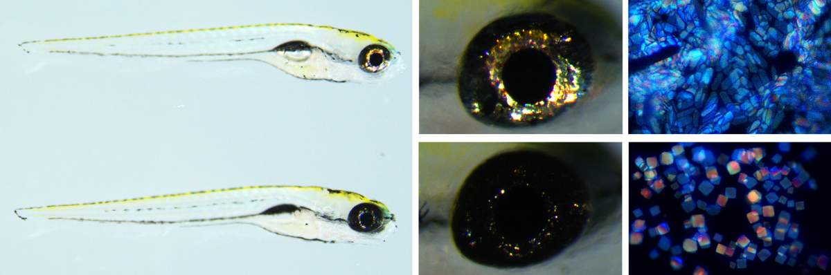
The researchers eventually put the mechanism to the test. They engineered a fish incapable of expressing an enzyme known as pnp4a, one of those needed to produce crystalline guanine. In a regular zebrafish, this enzyme is present in large quantities in the eye iridophores and in lower quantities in the skin iridophores. When the researchers engineered a fish incapable of expressing this enzyme, the crystals in its eyes were smaller in number and they changed shape from elongated to square, becoming similar to those seen in the fish's skin. The experiment confirmed that each group of cells has a unique enzyme balance and that when this balance is upset, there are changes in the cell's building blocks that lead to a change in the structure of its crystals.
"We have managed to reveal the whole story from beginning to end: from the genes that enable the fish to produce a wide variety of enzymes that have similar but different functions, through the unique expression level of each enzyme in the cells, to the way in which the presence of the enzymes determines the ratio between the different building blocks of the crystals - and therefore their characteristics," Gur summarizes. The researchers attribute their success in uncovering the entire mechanism to the interdisciplinary nature of the research team, which saw biologists working alongside experts in optics and biomaterials. Their findings not only further our understanding of nature and enable us to imitate its materials but also highlight the beauty and simplicity of nature: how two simple molecular structures produce such a wealth of complex biological functions.

Science Numbers
Crystals are formed in zebrafish around 44 hours after the eggs are fertilized. Each crystal-producing cell contains around 300 crystals, and each of them is made up of around 4 billion molecules of guanine and hypoxanthine.
Also participating in the study were: Olha Baiko, Dolev Brenman-Begin, Dr. Tali Lerer-Goldshtein, Dr. Zohar Eyal and Dr. Tsviya Olender from Weizmann's Molecular Genetics Department; Dr. Moshe Goldsmith from the Biomolecular Sciences Department; Yonghui Dong, Dr. Ziv Porat and Dr. Uwe Heinig from the Life Sciences Core Facilities Department; Dr. Sofya Lushchekina from the Brain Sciences Department; Dr. Smadar Levin-Zaidman, Dr. Iddo Pinkas and Dr. Neta Varsano from the Chemical Research Support Department; Sylvia Kaufmann and Dr. Rita Mateus from the Max Planck Institute; and Dr. Meital Kupervaser from the Nancy and Stephen Grand Israel National Center for Personalized Medicine.
Dvir Gur's research is supported by the Knell Family Center for Microbiology and the Henry Krenter Institute for Biomedical Imaging and Genomics.