A closer look at 50 years of imaging developments at EMBL
By Matthew Hartley, Christoph Müller, and Jan Ellenberg
In the late 1970s, John Kendrew, EMBL's first Director General, put Swiss biophysicist Jacques Dubochet in charge of the 'Laboratory for Electron Microscopy Applications'. The aim was to set up methods for carrying out electron microscopy at very, very low temperatures. The breakthrough came when the team figured out how to freeze samples quickly enough that the water in them would 'vitrify' rather than 'crystallise', a method that made it possible to observe biomolecules in solution at a resolution rarely achieved before.
Dubochet would go on to win the Nobel Prize in Chemistry in 2017, together with Joachim Frank and Richard Henderson, for the development of cryo-electron microscopy (cryo-EM). However, this is only one of many examples where technological advancement has progressed hand in hand with research at EMBL to push the field of bioimaging across new frontiers.
Reaching across fields with electron microscopy
The path to Dubochet's discovery had already been paved by the likes of Kevin Leonard, who joined EMBL as a group leader in 1975 and pioneered the use of electron microscopy in structural biology, setting up one of the first electron microscopy (EM) facilities at EMBL and investigating muscle structure and membrane proteins. After the Dubochet Group's breakthroughs in the 1980s, one of the first to realise cryo-EM's potential was Steve Fuller, who joined EMBL as a postdoc in 1981, before becoming a group leader in 1986 and later leading the structural biology programme. Fuller, through his work on the structures of the Semliki Forest Virus and HIV, was one of the key driving forces for cryo-EM development and application of computational image processing methods in biology.
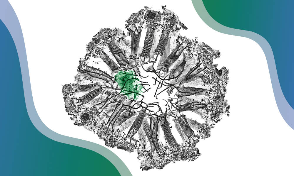
In the 1990s, cryo-EM group leaders at EMBL included Werner Kühlbrandt and later, Andreas Hönger and Bettina Böttcher. Kühlbrandt determined the high-resolution structure of the plant light-harvesting complex LHC-II by electron diffraction using 2D crystals, while Hönger applied helical reconstruction to the study of cytoskeletal dynamics. Böttcher, on the other hand, focused on membrane channel proteins that help the cell regulate its energy flows. During that time, the field of cryo-electron tomography (cryo-ET) - one can imagine it as something like a CT scan for cells - came into existence, and in the early 2000s, EMBL hired its first experts in cryo-ET - Achilleas Frangakis, John Briggs, and later Martin Beck.
Another electron microscopy method, volume EM (vEM), in which electron microscopy techniques are applied to 'large' volumes to generate a three-dimensional view of a cell, tissue, or an entire organism, was first introduced at EMBL in the early 2010s by the Electron Microscopy Core Facility (EMCF) and the group of Yannick Schwab . In recent years, this versatile technique has seen widespread use in many life science fields. In addition to pioneering the use of vEM for answering fundamental biological questions, EMBL has also been a leader in integrating vEM with other imaging technologies, especially 3D fluorescence microscopy and X-ray imaging.
Advanced electron microscopy techniques continue to be used by EMBL scientists to ask fundamental biological questions. Thanks to the 'resolution revolution' in cryo-EM - a term coined by EMBL alumni Werner Kühlbrandt to describe the technology leap caused by better detectors, improved software, and more stable microscopes - nowadays we can image molecular machines at quasi-atomic resolution using single-particle cryo-EM. As another example, Julia Mahamid's group at EMBL Heidelberg uses cryo-ET to observe the inner workings of a tiny pathogenic bacterium, Mycoplasma pneumoniae. Today, the EMBL Imaging Centre offers cryo-EM, cryo-ET, and vEM as services to researchers across the world, helping them investigate a plethora of biological systems.
Pushing the boundaries of light microscopy
In parallel to these developments in electron microscopy, advances in light microscopy were also growing at EMBL during those early years. An initial challenge was to be able to visualise cells in three dimensions, as traditional fluorescence microscopy could only view one plane in focus and blurred the remaining volume of the cell. Here, Ernst Stelzer at EMBL was key to introducing confocal microscopy to cell biology in the 1980s and to developing one of the first highly compact confocal microscopes that later became the basis for commercialisation by Carl Zeiss.
Another longstanding challenge in optical microscopy (and one of the drivers behind the development of electron microscopy) had been the diffraction limit - a physical principle which imposes a hard limit on the resolution achievable by light microscopes - around 250 nm. However, by the 1990s, scientists had already begun coming up with ingenious methods to get around this obstacle, ushering in the field of super-resolution microscopy.
One of the leading figures in this area was EMBL alumnus and later Nobel awardee Stefan Hell, who would go on to develop stimulated emission depletion (STED) microscopy in 1994, a super-resolution method which uses selective depletion of fluorophores to allow imaging at resolutions below the diffraction limit. STED allowed scientists to distinguish molecules placed only 70-90nm apart, which would be impossible in traditional optical microscopy.
Researchers at EMBL have subsequently used STED as well as other super-resolution techniques to study cellular structures in extraordinary detail and use these observations to reach insights into how living systems function at the molecular level. 2017 saw the introduction of MINFLUX, a super-resolution microscopy technique that could resolve molecules only 6 nanometres apart. The EMBL Imaging Centre was the first open-access facility to host a MINFLUX set-up and offer it as a service to scientists in Europe and globally.
While superresolution microscopy allows us to view subcellular structures and large molecular machines with incredible resolution and specificity, light sheet microscopy - a second pioneering technique to come out of EMBL - allows scientists to image and monitor live biological samples in three dimensions. The key contributors to its development included Ernst Stelzer, Jan Ellenberg, and Lars Hufnagel, and this technology also gave rise to Luxendo, one of EMBL's spin-off companies that focuses on developing and marketing state-of-the-art light-sheet microscopes and is now part of Bruker.
Scientists at EMBL today continue to experiment with the limits of light microscopy, using it in new and innovative ways to obtain novel biological insights. The team of Jonas Ries, who recently moved from EMBL Heidelberg to Max Perutz Labs at the University of Vienna, has been instrumental in developing new super-resolution microscopy techniques and using them to study cell biology, including the mechanisms of cellular endocytosis. The group of Robert Prevedel , on the other hand, has made new strides into using a kind of light microscopy called Brillouin microscopy to investigate the mechanical properties of cells, a development that was selected as one of The Guardian's '10 biggest science stories of 2022'.
Sharing innovations with the world
Advanced imaging techniques produce huge datasets, which must be properly analysed, shared, and archived. Here, a key role has been played by EMBL-EBI, which was established in 1992 and has played a major role in the bioinformatics revolution. In 2019, EMBL-EBI announced the launch of the BioImage Archive , a large-scale, free, publicly available and centralised data resource to host reference imaging data. The Archive builds on a long foundation of EMBL-hosted public archives for molecular and imaging data, which includes EMDB (the Electron Microscopy Databank, 2003) and EMPIAR (Electron Microscopy Public Image Archive, 2013).
In addition to working on storing and archiving bioimaging data, EMBL research and service groups have also been instrumental in finding new ways of making such data easily accessible, analysable, and visualisable by groups across the world. This has relied on the computational and experimental expertise of groups such as those currently led by Jan Ellenberg , Anna Kreshuk , Christian Tischer , Yannick Schwab , and Detlev Arendt .
Across all these developments, one of EMBL's key strengths has been its ability to bring together scientists or engineers interested in technology development and biologists interested in answering fundamental questions about how life works. The work done by the latter often gives rise to complex biological samples that present unique challenges for imaging. At the same time, the atmosphere of innovation ensures that there are always those willing to work with these challenging samples to develop solutions.
In addition to aiding its own biological research, EMBL's mission has also always been to improve and expand access to advanced imaging technologies to the entire scientific community in Europe and beyond. In addition to serving EMBL scientists, its Advanced Light Microscopy Facility and the Electron Microscopy Core Facility , headed by Rainer Pepperkok and Yannick Schwab , have together helped hundreds of users from across the world use microscopy to deepen our collective understanding of living systems.
In 2021, the EMBL Imaging Centre (EMBL IC) , headed by Jan Ellenberg and Christoph Müller , as an external user centre for the most advanced microscopy technologies opened its doors, driven by both the increase in sophistication of and the rising demand for access to high-end imaging technologies in its member states. The EMBL IC provides services of imaging techniques across scales, increasingly integrating light and electron microscopy and deploying them to study life in context. It aims to bring together technology development and service provision, helping develop novel correlative workflows and applications and enhancing close interaction with industry to allow new technological developments to become immediately available to the user community.
In this regard, EMBL has also been the driving force for Euro-BioImaging, the European landmark research infrastructure for biological and biomedical imaging, coordinating it from its first planning phase in 2008 until the establishment of the Euro-BioImaging ERIC in 2019, with Jan Ellenberg serving as the coordinator for biological imaging. EMBL has also always been at the forefront of providing training in high-end imaging techniques through workshops, courses, and conferences to the global scientific community, with specialised training offerings for everyone from undergraduates to established researchers and technicians.
The road ahead
It is exciting to speculate about the new roads that we will be travelling in bioimaging in the next fifty years and beyond. In the near future, we will likely see further major advances in biological imaging modalities and techniques, as well as their applications to unique biological questions. One area in which we are already seeing rapid advances is in the integration of imaging with omics approaches. By correlating imaging with gene and protein expression data on a whole-cell or tissue scale, the new fields of spatial transcriptomics and spatial proteomics allow to obtain a more comprehensive view of living systems.
Other areas of rapid development are large sample volume EM and intravital imaging, the latter being particularly important for fields like developmental biology. Finally, we are moving from an era of primarily focusing our imaging on model organisms to imaging diverse samples, including those collected from the environment, like during the TREC expedition. This, along with integrating imaging with other techniques like X-ray imaging and nano-beam imaging, are the objectives of the pan-European consortium IMAGINE, again coordinated by EMBL, that builds the bridging technologies that allow us to connect these modalities across Europe's research infrastructures. In the future, these new developments will allow us to reach incredible molecular resolutions in situ, helping us achieve unprecedented new insights into the inner workings of living systems.
Learn more about EMBL's contribution to life science research and services in our 50th anniversary commemorative publication.


