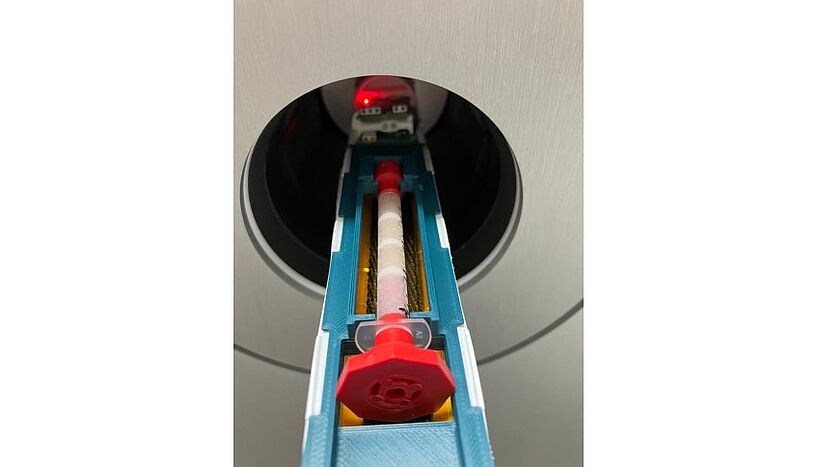
Fig. 1: The reconstructed 3D cell tissue is subjected to a PET scan.
Innovative system enables efficient evaluation of future cancer diagnostics
An FFG-funded consortium of Austrian research groups from the University of Vienna, MedUni Vienna and Technikum Wien together with company partner DOC Medikus GmbH has developed an innovative bioanalytical test system for radiopharmaceutical drug candidates for cancer diagnosis and therapy. It does not require any animal testing at all and enables automated, fast and highly precise analyses. The new method was presented in detail in the renowned Journal of Nuclear Medicine.
New drugs and diagnostic methods should be safe and ideally available quickly - but the preclinical test phase in particular often slows down rapid progress due to the high level of resources required. The development of radioactive marker substances ("radiotracers") in particular, which make physiological and pathological processes in the body visible and can be used in cancer diagnostics, for example, requires time-consuming and cost-intensive tests that have often been based on animal experiments to date. However, these are not only ethically controversial, but often provide results that are not transferable to the human body. An interdisciplinary research team from the University of Vienna, the University of Applied Sciences Technikum Wien, MedUni Vienna and DOC Medikus GmbH has developed an innovative solution: a bioanalytical test system that uses human cells on a silk matrix to test active ingredients under realistic conditions - faster, more precisely and without animal testing.
Ideas in flow
The already patented process combines chromatographic principles (separation of substances based on their interactions with a stationary and a mobile phase) with a dynamic 3D cell culture. At the heart of the process is a stationary phase made of biocompatible silk fibroin sponges, which act as an artificial scaffold to immobilize human cells in a three-dimensional structure. A special pump system continuously supplies the cells with nutrients, simulating realistic conditions in human tissue, while the radiopharmaceutical agents are applied and observed in real-time using imaging techniques (µPET/CT, positron emission tomography/computed tomography). This enables a parallel evaluation of radiotracer binding and cellular biochemical processes. First author Verena Pichler from the Department of Pharmaceutical Sciences at the University of Vienna explains: "With our method, we are not only creating an alternative to animal testing, but can also make the development of new radioactive marker substances much more efficient. Our aim is to raise diagnostics and therapy to a new level and improve ethical standards at the same time."
Relevance for practice
The new system enables a precise evaluation of the binding properties of radioactive marker substances to be tested, their target accuracy and possible side effects. The use of silk fibroin offers considerable advantages due to its radiation stability and its proven application in cell culture. The introduction of frits (sieve-like partitions) between the sponges reduces cell migration and improves the reproducibility of results. Important factors such as the distribution of the radiation dose and the supply of nutrients to the cells can thus be precisely controlled. Particular attention was paid to the automation and standardization of the processes in order to make the processing of the radioactive substances safe and efficient. The new method complies with the recommendations of the 3R principle ("reduce, refine, replace") and the FDA's Critical Path Initiative. It has the potential to significantly reduce animal testing, accelerate the development of radiopharmaceuticals and minimize radiation exposure for personnel. This groundbreaking technology could set new standards in preclinical radiopharmacy - for more sustainable and efficient drug development.
The Spheriograph project to develop the innovative 3D cell culture system was funded by the Austrian Research Promotion Agency (FFG) as part of a bridge project.
Original publication
Verena Pichler, Verena Schwingenschlögl-Maisetschläger, Irem Duman, Xavier Monforte, Stefanie Ponti, Lukas Zimmermann, Elma Joldic, Monika Dumanic, Chrysoula Vraka, Marcus Hacker, Christian Kraule, Andreas Herbert Teuschl-Woller.
Bioanalytical Hybrid System Merging 3D Cell Culture and Chromatographic Precision for Unprecedented Preclinical Insights in Molecular Imaging. In Journal of Nuclear Medicine.
Pictures
Fig. 1: The reconstructed 3D cell tissue is subjected to a PET scan. C: Verena Pichler
Fig. 2: Assembling the system is particularly user-friendly. C: Verena Schwingenschlögl-Maisetschläger
Fig. 3: Regular monitoring of the novel cell system enables precise analysis of drugs and their mode of action. C: Verena Schwingenschlögl-Maisetschläger


