Three-dimensional imaging has become an essential tool for Australian palaeontologists to more accurately analyse and interpret the life histories and relationships between dinosaurs.
The latest example is groundbreaking research published today in the Journal of Vertebrate Paleontology by the Museums Victoria Research Institute and Monash University that unveiled a landmark discovery - fossils of the world's oldest known megaraptorid and the first evidence of carcharodontosaurs in Australia.
Researchers used the Imaging and Medical beamline (IMBL) at ANSTO's Australian Synchrotron in their study to acquire detailed images of the fossils.
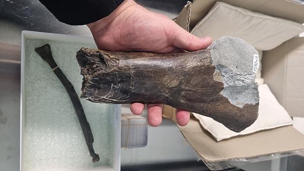
One of the five theropod fossils discovered along Victoria's coastline, a shin bone belonging to one of Australia's largest known carnivorous dinosaurs, was scanned using IMBL.
"This sample challenged the limits of the beamline's physical capability- it needed the highest energy X-rays of the brightest X-source in the southern hemisphere to peer inside the bone," said Dr Jospeh Bevitt, a co-author on the publication, who assisted the team with measurements.
"From this imaging study, we were peer inside the fossil and determine that this bone was from an adult megaraptorid - a real apex predator of its time".
Dr Bevitt assists palaeontologists from across the globe with the imaging of fossils using the Neutron tomography instrument Dingo at the Australian Centre for Neutron Scattering, and the Imaging and Medical beamline at the Australian Synchrotron.


