WashU Researchers Shine Light On Amyloid Architecture
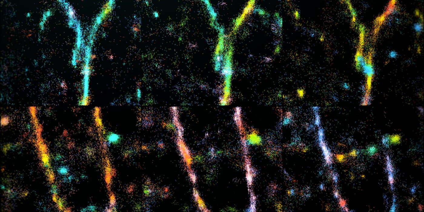
Time-lapse single-molecule orientation-localization microscopy (SMOLM) of amyloid beta (Aβ42) fibrils undergoing remodeling, taken over 10 minutes, shown as a multi-colored mosaic. Transiently binding and flashing Nile blue (NB) molecules enable the nanoscale architectures of growing and decaying regions in fibrils to be visualized as these multicolored forks. (Image: Lew lab)
/Public Release. This material from the originating organization/author(s) might be of the point-in-time nature, and edited for clarity, style and length. Mirage.News does not take institutional positions or sides, and all views, positions, and conclusions expressed herein are solely those of the author(s).View in full here.