18 April 2024
Experiments with neutron and X-ray radiation provide molecular insights into the iron metabolism of marine bacteria
The cyanobacterium Prochlorococcus is the smallest and most abundant photosynthetic organism in the world. And it has another remarkable property: in the course of evolution, it has found a genetically particularly efficient way to absorb and store vital iron. It has a multifunctional protein that can bind both divalent (Fe2+) and trivalent (Fe3+) iron ions thanks to a molecular switch. Using neutron, X-ray, and synchrotron experiments, an international team of researchers involving employees of Forschungszentrum Jülich has visualized the molecular mechanism for the first time.
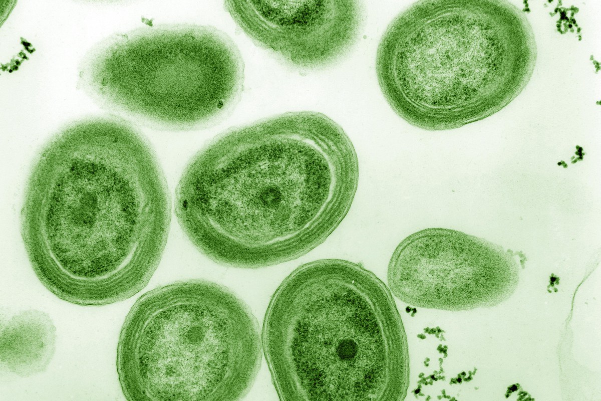
The sea is the largest ecosystem on Earth. Organisms living in the sea, which carry out photosynthesis, produce around half of the oxygen on Earth. At the same time, they are an important carbon sink in the fight against climate change. Every year, they bind a total of 45 gigatonnes of carbon. Photosynthetic bacteria are one of the main players here, and the cyanobacterium Prochlorococcus holds several records in this field: with a diameter of half a micrometre - the equivalent of half a millionth of a metre - it is the smallest photosynthetic organism on Earth. It produces four gigatonnes of sequestered carbon per year, which corresponds to the annual net primary production of global agriculture. Its photosynthetic activity, however, depends on iron, which is only available to a limited extent in the ocean.
Biologically relevant iron can occur in nature in two different oxidation states, as divalent or trivalent iron ions, Fe2+ and Fe3+ - i.e. as a charged atom that has released two or three electrons. Most organisms, including cyanobacteria, usually have two different proteins to absorb these different oxidation states. This is different for Prochlorococcus, whichcan bind both forms of iron with a single protein.
The researchers suspect that this dual function is an important reason for the ecological success and is linked to the compact genome of Prochlorococcus. As this marine bacterium is so tiny, it only has a greatly reduced genome and therefore has to manage with a limited number of genes that could be translated into different proteins.
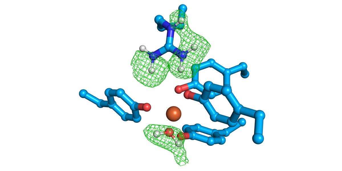
An international research group headed by Prof. Dr. Ivo Tews from the University of Southampton has now been able to demonstrate for the first time the mechanism by which Prochlorococcus absorbs the two different forms of iron. The researchers discovered a molecular switch that enables the bacterium to switch back and forth between binding divalent and trivalent iron ions. The "FutA protein" also has another function. In the bacterium, it is not only responsible for the uptake, but also for the protection of the iron that has already been absorbed.
Combination of X-rays, neutrons, and spectroscopy
The discovery was made possible by a novel combination of experiments. The researchers used X-rays, neutron beams, and light absorption to understand the iron binding process. They also used X-rays to switch between the two forms of iron. In several experiments carried out at the Diamond Light Source and also at an X-ray laser facility in Japan, the researchers determined the position of the atoms - with the exception of the hydrogen atoms. Their arrangement could only be made visible with the help of neutron beam experiments.
Experiments with neutron beams at the research neutron source Heinz Maier-Leibnitz (FRM II) in Garching thus helped to understand the state of charge of the iron.
"The BIODIFF diffractometer, which Forschungszentrum Jülich operates together with the Technical University of Munich, is designed for determining the position of hydrogen atoms in such large molecules," explains Dr. Tobias Schrader from the Jülich Centre for Neutron Science (JCNS). Based on this, the researchers calculated the charges of the amino acid side chains in the protein that are located around the iron. These in turn allow conclusions to be drawn about the charge of the iron.
"Using a nuclear reactor to see the hydrogen atoms was really exciting for me. Even the hydrogen atoms in the water were clearly visible," says Rachel Bolton, doctoral student at the University of Southampton, who carried out the experiments in Garching.
Original publication
Rachel Bolton et al.
A redox switch allows binding of Fe(II) and Fe(III) ions in the cyanobacterial iron-binding protein FutA from Prochlorococcus
PNAS (2024), DOI: 10.1073/pnas.2308478121


